Tuesday, 8 October 2024


    Legal Considerations in Orthopaedic Practice


                               Dr. KS Dhillon


Informed Consent

Components 

When obtaining an informed consent the patient must be provided with all the information needed to make an informed decision. The patient must be told of the diagnosis or medical problem for which treatment is recommended. The patient also has to be told who will be performing the surgery. 

If the surgeon has tested positive for HIV/HBV/HCV the information has to be disclosed to the patient at the time of scheduling an "exposure-prone" procedure. The patient has to be told of the proposed treatment or procedure. This would include its purpose, duration, methods, and implements used, and the probability of success.

The origin of surgical implants should be discussed with the patient. This may have implications for their use based on a patient's religious background. 

In Hinduism, the use of bovine-derived implants should be discussed and in Judaism and Islam, the use of porcine-derived implants should be discussed.

The patient has to be told of all material risks of the procedure or treatment. The patient has to be told about any reasonable alternatives to the proposed procedure and of the risks of not being treated.

Special situations

Patient consent is not needed when communicating Health Information Portability and Accountability Act (HIPAA) protected information to other treating providers.

Institutional Review Board (IRB) approval for obtaining informed consent from patients enrolled in clinical trials is required. IRB approval is not required for quality improvement studies used for internal purposes only. 

The IRB aims to ensure the rights and welfare of human subjects participating in research.  

Informed consent for elective surgical procedures is best obtained in the office/clinic setting a few days before the scheduled procedure. 

In life-threatening injuries requiring surgical intervention but without available legal consent, the surgeon should confirm and document the necessity of care with a fellow orthopaedic surgeon or colleague. In non-life threatening injuries, consent must be obtained prior to surgical intervention. 

 When patients are non-consentable the legal guardians have the highest precedence. The "next of kin" precedence has been established to assist in determining the order of consent.  

Patient-physician relationship

Termination of care can be initiated by the physician with due process. The patient must be notified in writing the relationship will be terminated. A grace period of 30-45 days of continued care should be given to allow the patient to arrange for further treatment. Termination without a grace period is considered abandonment.


Physician Errors

Communication errors are the leading cause of wrong-side surgeries, medication errors, diagnostic delays, or loss to follow-up. These errors result in increased treatment costs, treatment delays, and complications. 

Crew resource management has been shown to improve communication and team dynamics. It has led to an improvement in patient safety and team morale. 

Wrong site surgery has to be prevented. The patient should be involved in identifying the correct side in the pre-operative area prior to induction. 

Pertinent imaging must be displayed in the operating room. The correct site with the surgeon's initials should be marked visibly in the surgical field. A time-out should be performed with the operating room team prior to the incision.

When wrong site surgery has been performed the error must be  

acknowledged with immediate discussions with the family. There is a need to apologize and accept responsibility. Blame must not be placed on others. 

Surgical errors can be prevented by having a surgical "time-out".

According to the Joint Commission on Accreditation of Healthcare Organizations (JCAHO), time out should include the following: 


  • identify the correct patient, site, and side

  • verify the correct procedure

All members of the team should be present for the time out.

WHO implementation of the surgical safety checklists began in 2009. It resulted in measurable improvements in surgical mortality, in-hospital complications, and adherence to surgical plan in OR crisis situations (e.g., massive hemorrhage, cardiac arrest). The surgeon is the most effective OR team member at reducing complications when using the surgical checklist and "time-out".

Medication prescribing errors are reduced when physicians use computerized order entry. 

Medical documentation errors are sometimes seen. Altering the medical record for any reason is illegal. No one has the authority to authorize a physician to alter the medical record. The errors can be noted and addendums can be added.

The second-opinion surgeon is ethically required to disclose the effect of medical errors on patient outcome. Only the patient can unilaterally decide to transfer care to a second surgeon. The surgeon is not ethically allowed to seek out transfer of care of a patient.


Litigation

In the USA medical liability lawsuits involving orthopaedic surgeons increased by 13% from 2003 to 2008. It is thought to be related to the aging population. Compared to other specialties, orthopaedic surgery has the 7th highest number of lawsuits. About 33% of all orthopaedic surgery claims result in payment to plaintiffs. The average cost of defending orthopaedic surgery claims is about USD 47,000. 

"Improper performance" makes up 45% of lawsuits. The most commonly associated procedures include:

  • open reduction of dislocation

  • closed reduction of fractures

  • operative procedures of joint structures (not including spinal fusion)

  • operative procedures on bones

  • operative procedures on cranial and peripheral nerves

The most commonly associated clinical diagnoses include:

  • osteoarthritis (21%)

  • disorder of joints, not including arthritis

  • fracture of femur


Legislation

In the USA there are several legislation that protect the patient.

1. Stark Law (1993)

A federal regulation that prohibits the self-referral of physicians to organizations with which they have a financial relationship. 

2. Patient Protection and Affordable Care Act (2010)

It provides numerous rights and protections that make health coverage fairer, easier to understand, and more affordable.

3. Physician Payments Sunshine Act (2010)

It requires the collection and reporting of financial relationships between physicians/teaching hospitals and businesses (manufacturers of drugs, devices, medical supplies). All payments of more than $10 must be reported to Centers for Medicare and Medicaid Services.


Physician Impairment

Physician impairment is defined as the inability or impending inability to practice according to accepted standards due to substance use, abuse, or dependency/addiction.

Surgeons who discover chemical impairment, dependence, or incompetence of a colleague or supervisor have a responsibility to ensure that the problem is identified and treated.


Medical Negligence

Medical negligence is the failure to provide the standard of health care resulting in medical injuries. A second-opinion physician has an ethical obligation, but not a legal obligation, to disclose if the standard of care has been breached by a treating physician. 

 Successful patient-plaintiff lawsuits for medical negligence require that all of the following 4 elements be alleged and proven in a court of law. 

Duty - Obligation to provide care that meets the professional standard of care, i.e. the same standard of care ordinarily executed by surgeons in the same medical specialty. 

Breach of duty - occurs when action or failure to act deviates from the standard of care.

Causation - established if it is demonstrated that failure to meet the standard of care was the direct cause of the patient’s injuries.

Damages - monies awarded as compensation for injuries sustained as the result of medical negligence.


Workers Compensation

Maximum medical improvement is reached when further restoration of function is no longer anticipated, allowing patients to settle their claim. 

Impairment occurs when there is a loss of function resulting from an anatomic or physiologic derangement. 

There is disability when there is limitation of an individual’s capacity to meet certain personal social or occupational demands.


Physician Employment

When the physician is employed as an independent contractor the employer influences the outcome. The contractor determines the methods and means of achieving the result of the work. The employer does not pay taxes, provide insurance, or offer retirement benefits. 

When the physician is employed the employer determines the result of the work and provides the means and methods for the result. The employer also provides resources and training. The employer pays taxes, provides insurance, and retirement benefits

Friday, 4 October 2024

                Osteopetrosis


                                DR KS Dhillon


Introduction

The term osteopetrosis is derived from the Greek language. In the Greek language ‘osteo’ means bone, and ‘petrosis,’ means stone. Hence, the disease is colloquially often referred to as “marble bone disease.” The disease was originally described by Dr. Albers-Schonberg, a German radiologist in 1904 (1). Increased bone density is the key radiographic finding. The increased density is secondary to osteoclast dysfunction. This leads to the affected bones being abnormally brittle (1). 

The term osteopetrosis encompasses a group of hereditary metabolic bone diseases. All these diseases detrimentally affect bone growth and remodeling leading to generalized osteosclerosis. With osteosclerosis, there is a potential for pathologic fractures, pancytopenia, and even cranial neuropathies and hepatosplenomegaly in severe cases (2-4).

There are 4 known disease forms. One is the malignant autosomal recessive form. It is not named malignant due to any relation to oncology. It is rather due to the degree of condition severity which is very severe and often leads to mortality in early childhood (5,6). The other is the intermediate autosomal recessive form. This usually becomes clinically significant during the first decade of life. Patients with this form often suffer pathologic fractures and progressive cranial nerve compression neuropathies. They however typically live into adulthood. For autosomal dominant osteopetrosis, there are two subclassifications, and these patients are often asymptomatic into adulthood. The type I does not have increased fracture risk. It presents with isolated osteosclerotic thickening of the cranial vault. Patients with type II usually present in adulthood with pathologic fracture, anemia, or early arthritis (7).


Etiology

Genetic studies have found that this disease of osteoclastic dysfunction has an association with at least 8 gene mutations (3). Six of these 8 genes are associated with a malignant, autosomal recessive form of the disease. Loss of function mutations in CLCN7, OSTM1, TCIRG1, PLEKHM1, AND SNX10 lead to an osteoclast rich version of autosomal recessive osteopetrosis. In this version, there are a lot of osteoclasts. However, these osteoclasts are unable to resorb bone effectively due to defective ruffled border formation. Loss of function mutations in TNFRSF11A and TNFSF11 leads to disrupted osteoclast development and osteoclast poor osteopetrosis (8).

Intermediate autosomal recessive osteopetrosis is the result of a loss of function mutation in CAII. This gene is responsible for the production of the carbonic anhydrase II protein (3).

Autosomal dominant osteopetrosis results from dysfunction of chloride channel 7 secondary to a dominant-negative mutation of CLCN7 (3).


Epidemiology

The autosomal recessive form of the disease is far less common than the autosomal dominant form. The autosomal recessive form occurs in about 1 out of every 250,000 births. In Costa Rica, the incidence is significantly higher, with a rate of approximately 3.4 out of every 100,000 births (5). 

The frequency of the autosomal dominant form of the disease is about 1:20,000 (9).


Pathophysiology

Bone is in a dynamic state. It is dependent upon a healthy balance between osteoclast-mediated resorption and osteoblast-mediated deposition of bone. In osteopetrosis, there is defective osteoclast development or function that leads to a disruption in normal bone homeostasis (8,1). Osteoclasts with defective proton pumps, chloride channels, or carbonic anhydrase II proteins are unable to resorb bone effectively. This leads to the formation of unorganized, overly dense bone that is prone to fractures.


Histopathology

Histological examination of bone in patients with osteopetrosis usually shows empty lacunae with plugged Haversian canals, calcified cartilage dispersed within bony trabeculae, and defective osteoclasts that lack a clear zone and ruffled border. These clear zone and ruffled border structures are classical findings in osteoclasts undergoing active resorption, and their absence corresponds with the findings of osteopetrosis (10).


Clinical Presentation

The history and physical examination findings differ drastically depending on the type of osteopetrosis the patient has. The malignant, autosomal recessive osteopetrosis presents in infants within a few months following birth. They present with symptoms that include frequent infections, abnormal bruising, and bleeding abnormalities. These symptoms occur due to the bone that is no longer being correctly resorbed by osteoclasts and is consequently encroaching into the medullary space (3). Pathologic fractures are common among these patients. There are additional symptoms that can occur. These include macrocephaly, nasal congestion, hepatosplenomegaly, and dental abscess or osteomyelitis of the mandible (2). Diseased bone tends to narrow cranial nerve foramina in children leading to progressive deafness and blindness along with possible facial palsies. The most frequently involved cranial nerve is the optic nerve. This is followed by the auditory nerve, trigeminal nerve, and the facial nerve (11,1). 

The history and physical examination of patients with intermediate autosomal recessive osteopetrosis are very variable. Symptoms are similar but are not as severe and do not present as early as the symptoms present in the malignant form of the autosomal recessive disease. Since this variety of osteopetrosis can be secondary to carbonic anhydrase II dysfunction, renal tubular acidosis can be present in these patients (12).

Type 1 autosomal dominant osteopetrosis usually has a very mild clinical presentation. Unlike other forms of osteopetrosis, type 1 autosomal dominant osteopetrosis is due to an error in increased bony formation rather than a defect in osteoclast function (13,14). Hence these patients do not have increased fracture risk as in other patients with osteopetrosis (9). In this form, osteosclerosis is most focused in the cranial vault, and cranial nerve compression neuropathies are common (15). 

Type 2 autosomal dominant osteopetrosis is the most common form that doctors treat, and it has a very heterogeneous course. Most patients with this form of the disease lead a relatively normal life. They generally have a normal life span, physique, and overall health. Patients often discover that they have this disease after they present for evaluation of a pathological fracture or early-onset osteoarthritis. Fractures occur in about 4 out of 5  patients. The average number of fractures per affected individual is three. Most pathological fractures occur in the femur (16). Arthritis can occur in a variety of locations. The hip is the most common location, with about 50% of patients having early-onset hip pain (16). Fatigue due to anemia and cranial nerve neuropathies can affect these patients as well but occurs less frequently than in other forms of the disease. Optic and/or auditory nerve damage occurs in approximately 1 out of 20 patients (17).


Evaluation

Osteopetrosis is usually diagnosed based on the presence of typical clinical and radiographic findings of the disease. The radiographs usually show diffuse osteosclerosis throughout the skeleton with a “marble bone” appearance. There is increased cortical thickness with associated decrease in medullary canal diameter. There will be an “Erlenmeyer flask” deformity at the metaphyses of long bones, especially at the proximal humerus and the distal femur. A “bone-in-bone” appearance is frequently noted in the bones of the spine or phalanges of the hand. “Rugger jersey spine” is another axial skeleton radiographic finding. It can occur due to excessive sclerosis of the vertebral endplates (1).

If radiographic and clinical findings do not lead to a diagnosis, laboratory findings of increased creatinine kinase BB and tartrate-resistant acid phosphatase can help in the diagnosis (18). Genetic testing can also be done to evaluate the presence of the gene mutations associated with the condition.


Management

Treatment of patients with osteopetrosis should be tailored to the individual patient. Treatment is predominantly supportive. There is no known cure. Interprofessional care and surveillance are the mainstays of treatment. 

Fractures and arthritis associated with osteopetrosis are treated by orthopedic surgeons. Fracture treatment, and arthroplasty in these patients can frequently be associated with the following complications: delayed union, non-union, and osteomyelitis (19).

Routine ophthalmologic evaluation is needed since cranial nerve compression neuropathies are common. The optic nerve is frequently involved. In some patients, surgical decompression of the optic nerve may be required to preserve eyesight (20).

Routine dental evaluation is needed in these patients to look for complications such as cysts, abscesses, and osteomyelitis that can occur due to altered bony anatomy of the mandible (1).

Bone marrow transplantation of hematopoietic stem cells (HSC) is reserved for the malignant, autosomal recessive form of osteopetrosis. There are risk of rejection and other possible complications. HSC therapy from HLA-matched donors does not necessarily reverse disease complications. It has been found to have a 73% 5-year disease-free survival (21).

In some patients who are unfit for bone marrow transplantation, interferon-gamma 1b therapy has been used. It can also be used as bridging therapy until HSC therapy can be used. It increases immune function and bone resorption (22). High-dose calcitriol has also been used to stimulate host osteoclasts (23).


Differential Diagnosis

Osteopetrosis is a disease of primary bone sclerosis. Many conditions can lead to similar osteosclerosis. The following conditions must be kept in mind when evaluating a patient with osteosclerotic bone seen on radiographic evaluation:

  • Fluorosis

  • Myelofibrosis

  • Beryllium, lead, and bismuth poisoning

  • Paget disease

  • Cancer (lymphoma or osteoblastic bony metastases)


Prognosis

Without successful bone marrow transplantation treatment, the malignant autosomal recessive form of osteopetrosis is frequently fatal in the first years of life. Many patients undergo multiple attempts at bone marrow transplants without long-lasting success. The other disease forms usually allow patients to live into adulthood. The autosomal dominant form of the disease has very little effect on life span and overall health.


Complications

Complications include refracture in patients with pathologic fractures. Refractures occur due to the brittle nature of sclerotic bone. Hardware failure is also a common problem in patients with osteopetrosis who require fracture fixation. Peri-prosthetic fractures can further complicate care. These patients may require specialized orthopedic tools intraoperatively since the hard, brittle bone can cause failure in some tools. Bone infections are common due to disrupted bone vascularity. Malunion and non-union of pathologic fractures can occur.


Conclusion

Treatment for patients with osteopetrosis is best delivered by an interprofessional team that consists of pharmacists, nurses, physical therapists, and physicians. This team design allows for complex patients to have patient-centered care that leads to improved outcomes.

Diagnosis is usually made by clinical and radiographic assessment. These patients will require hospitalization at some point in their life to treat complications that occur. 

These patients will invariably require orthopedic care. Neurosurgeons may be required to treat cranial compression neuropathies. Ophthalmologists and dentists are required to provide maintenance and surveillance care for at-risk patients. Care for these patients with osteopetrosis is truly a team effort. 


References

  1. Stark Z, Savarirayan R. Osteopetrosis. Orphanet J Rare Dis. 2009 Feb 20;4:5.

  2. Reddy Mh R. Osteopetrosis (Marble Bone Disease): A Rare Disease in Children. Int J Clin Pediatr Dent. 2011 Sep-Dec;4(3):232-4. 

  3. Coudert AE, de Vernejoul MC, Muraca M, Del Fattore A. Osteopetrosis and its relevance for the discovery of new functions associated with the skeleton. Int J Endocrinol. 2015;2015:372156. 

  4. de Vernejoul MC, Bénichou O. Human osteopetrosis and other sclerosing disorders: recent genetic developments. Calcif Tissue Int. 2001 Jul;69(1):1-6. 

  5. Loría-Cortés R, Quesada-Calvo E, Cordero-Chaverri C. Osteopetrosis in children: a report of 26 cases. J Pediatr. 1977 Jul;91(1):43-7.

  6. Beighton P, Horan F, Hamersma H. A review of the osteopetroses. Postgrad Med J. 1977 Aug;53(622):507-16.

  7. de Baat P, Heijboer MP, de Baat C. [Osteopetrosis. Classification, etiology, treatment options and implications for oral health]. Ned Tijdschr Tandheelkd. 2005 Dec;112(12):497-503.

  8. Sobacchi C, Schulz A, Coxon FP, Villa A, Helfrich MH. Osteopetrosis: genetics, treatment and new insights into osteoclast function. Nat Rev Endocrinol. 2013 Sep;9(9):522-36.

  9. Bollerslev J, Andersen PE. Radiological, biochemical and hereditary evidence of two types of autosomal dominant osteopetrosis. Bone. 1988;9(1):7-13. 

  10. Shapiro F, Glimcher MJ, Holtrop ME, Tashjian AH, Brickley-Parsons D, Kenzora JE. Human osteopetrosis: a histological, ultrastructural, and biochemical study. J Bone Joint Surg Am. 1980 Apr;62(3):384-99. 

  11. Dozier TS, Duncan IM, Klein AJ, Lambert PR, Key LL. Otologic manifestations of malignant osteopetrosis. Otol Neurotol. 2005 Jul;26(4):762-6. 

  12. Sly WS, Hewett-Emmett D, Whyte MP, Yu YS, Tashian RE. Carbonic anhydrase II deficiency identified as the primary defect in the autosomal recessive syndrome of osteopetrosis with renal tubular acidosis and cerebral calcification. Proc Natl Acad Sci U S A. 1983 May;80(9):2752-6. 

  13. Henriksen K, Gram J, Høegh-Andersen P, Jemtland R, Ueland T, Dziegiel MH, Schaller S, Bollerslev J, Karsdal MA. Osteoclasts from patients with autosomal dominant osteopetrosis type I caused by a T253I mutation in low-density lipoprotein receptor-related protein 5 are normal in vitro, but have decreased resorption capacity in vivo. Am J Pathol. 2005 Nov;167(5):1341-8. 

  14. Van Hul E, Gram J, Bollerslev J, Van Wesenbeeck L, Mathysen D, Andersen PE, Vanhoenacker F, Van Hul W. Localization of the gene causing autosomal dominant osteopetrosis type I to chromosome 11q12-13. J Bone Miner Res. 2002 Jun;17(6):1111-7. 

  15. Oğütcen-Toller M, Tek M, Sener I, Bereket C, Inal S, Ozden B. Intractable bimaxillary osteomyelitis in osteopetrosis: review of the literature and current therapy. J Oral Maxillofac Surg. 2010 Jan;68(1):167-75.

  16. Bénichou OD, Laredo JD, de Vernejoul MC. Type II autosomal dominant osteopetrosis (Albers-Schönberg disease): clinical and radiological manifestations in 42 patients. Bone. 2000 Jan;26(1):87-93. 

  17. de Vernejoul MC, Kornak U. Heritable sclerosing bone disorders: presentation and new molecular mechanisms. Ann N Y Acad Sci. 2010 Mar;1192:269-77.

  18. Waguespack SG, Hui SL, White KE, Buckwalter KA, Econs MJ. Measurement of tartrate-resistant acid phosphatase and the brain isoenzyme of creatine kinase accurately diagnoses type II autosomal dominant osteopetrosis but does not identify gene carriers. J Clin Endocrinol Metab. 2002 May;87(5):2212-7.

  19. Landa J, Margolis N, Di Cesare P. Orthopaedic management of the patient with osteopetrosis. J Am Acad Orthop Surg. 2007 Nov;15(11):654-62. 

  20. Hwang JM, Kim IO, Wang KC. Complete visual recovery in osteopetrosis by early optic nerve decompression. Pediatr Neurosurg. 2000 Dec;33(6):328-32.

  21. Driessen GJ, Gerritsen EJ, Fischer A, Fasth A, Hop WC, Veys P, Porta F, Cant A, Steward CG, Vossen JM, Uckan D, Friedrich W. Long-term outcome of haematopoietic stem cell transplantation in autosomal recessive osteopetrosis: an EBMT report. Bone Marrow Transplant. 2003 Oct;32(7):657-63.

  22. Key LL, Ries WL, Rodriguiz RM, Hatcher HC. Recombinant human interferon gamma therapy for osteopetrosis. J Pediatr. 1992 Jul;121(1):119-24. 

  23. Kocher MS, Kasser JR. Osteopetrosis. Am J Orthop (Belle Mead NJ). 2003 May;32(5):222-8.

Monday, 30 September 2024

       Ollier disease

                                     Dr. KS Dhillon


Introduction

Enchondromas are common intraosseous, usually benign cartilaginous tumors. They develop close to the growth plate cartilage. When multiple enchondromas are present, the condition is called enchondromatosis. Enchondromatosis is also known as Ollier disease. The prevalence of Ollier disease is estimated to be 1 in 100,000 individuals. Clinical manifestations usually appear in the first decade of life. In Ollier disease, there is an asymmetric distribution of cartilage lesions. These lesions can be extremely variable in terms of size, number, location, age of onset, and requirement for surgery. Enchondromas can produce skeletal deformities and limb-length discrepancies. They have the potential risk for malignant change to chondrosarcoma. When multiple enchondromatosis is associated with soft tissue hemangiomas, the condition is known as Maffucci syndrome. Ollier disease and Maffucci syndrome occur in isolated patients. These conditions are not familial. It is uncertain whether the disorder is caused by a single gene defect or by combinations of germ-line and/or somatic mutations. The diagnosis is made by clinical and conventional radiological evaluations. Histological analysis has a limited role. It is mainly used if malignancy is suspected. Medical treatment has no role in the management of enchondromatosis. Surgery is usually indicated when there are complications such as pathological fractures, growth defects, and malignant transformation. It is difficult to assess the prognosis for Ollier disease. Forms with an early onset appear more severe. There is a risk of malignant transformation of enchondromas into chondrosarcomas in patients with Ollier disease.


Definition

Enchondromas are common benign cartilage tumors. They develop in the metaphyses and may become incorporated into the diaphyses of long tubular bones, in close proximity to growth plate cartilage (1-3). They are usually asymptomatic. Enchondromatosis or Ollier disease is defined by the presence of multiple enchondromas with an asymmetric distribution of cartilage lesions that can be very variable in terms of number, size, location, and age of onset (4).

Multiple enchondromatosis associated with soft tissue hemangiomas is known as Maffucci syndrome.


Epidemiology

It is the 2nd most common benign cartilage lesion. Osteochondroma is the most common. The male-to-female ratio is 1:1. It is most common in 20-50 year old individuals. It is usually found in the medullary cavity of the diaphysis or the metaphysis. The most common location is the hand (60%). It is more common in the hand as compared to the feet. The most common primary bone tumor in the hand is the enchondroma. Other locations include the distal femur (20%), proximal humerus (10%), and the tibia.


Clinical presentation

Ollier disease usually presents in the first decade of life. Clinical presentation usually starts with the appearance of palpable bony masses on a finger or toe, asymmetric shortening of an extremity, limp, osseous deformities with or without pathologic fractures (1-3). Physical examination usually shows visible masses embedded within phalanges, metacarpal, and metatarsal bones. Enchondromas usually affect long tubular bones, particularly the femur, the tibia, and/or the fibula. Flat bones, especially the pelvis, can also be affected. Multiple bones can be affected and the lesions are usually asymmetrically distributed, predominantly affecting one side of the body. Affected bones are quite often deformed and shortened. Bone shortening may be the only clinical sign of the disease. Bone shortening is often associated with bone bending and curving. This can lead to limitations in joint movements. Forearm deformities that are frequently encountered are similar to those observed in hereditary multiple exostosis (HME). The trunk is usually not affected, except for rib enchondromas and scoliosis which results from pelvis imbalance. In children, the lesions are subjected to pathologic fractures.

Clinical forms

Enchondromatosis has been recognized for a long time. Ollier at the end of the 19th century emphasized the asymmetrical and random distribution of enchondromas. Some authors have distinguished two subtypes of enchondromatosis i.e. enchondromatosis and Ollier disease. The first subtype affects mostly men. In this type, enchondromas are located mainly at the extremities and appear to be transmitted in an autosomal dominant fashion (5). The second form mainly affects women. In this subtype, there is sporadic unilateral distribution of enchondromas. The basis for this classification into two forms is not supported by a thorough analysis of available data. When multiple enchondromas are associated with hemangiomas it is referred to as Maffucci syndrome. Gabos and Bowen recently reported a previously unreported form in which there is extensive involvement of the epiphyseal and metaphyseal regions of long bones of the lower extremity (6).


Radiography

Enchondromas are most likely present at birth although they are rarely observed at birth. 

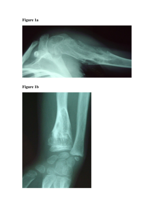
X-rays usually show multiple, radiolucent, homogenous lesions with an oval or elongated shape and a slightly thickened bony margin (fig 1a and 1b) (1-3). The lesions run parallel to the long bone axis. The lesions usually calcify with time. They become diffusely punctated or stippled, and a light trabeculation is usually visible. Enchondromas are frequently present as clusters. This leads to the metaphyseal widening. When they are localized at the bone border, they produce a typical notch-like image. A delay in bone age (average 0.6 +/- 1.3 years), has been reported in children with Ollier disease (7).

Enchondromas are usually localized in the metaphysis of long bones and in the small bones of the feet and hands. Initially, they are localized close to the growth plate cartilage. Later they migrate progressively towards the diaphysis. There may be irregularities in the epiphyseal region next to an affected metaphysis (1,6).


There is irregular distribution of the lesions. They can be localized to one limb, or limited to one half of the body. Though the lesions are limited largely to one side of the body, one or two enchondromas are frequently present on the other side, especially in the hand bones. When the lesions are distributed over the entire body, one side is usually more affected. The lesions in the hand almost never affect the metacarpal bones and phalanges.

Enchondromas can produce severe growth abnormalities. The growth abnormalities can be more severe than those observed in multiple exostosis. The affected diaphysis is short and massively enlarged. There may be bending close to the metaphysis. Ulnar shortening is usually more than shortening of the radius. The fingers are often of irregular sizes. Evidence of pathological fractures may be present.

Evidence of malignant transformation should be sought as it is a major complication of enchondromatosis. Signs of malignant transformation include extension of the tumor into soft tissues, cortical erosion, and irregularity or indistinctness of the surface of the tumour. Enchondromas are well circumscribed, chondrosarcomas on the other hand show poor demarcation. In differentiating enchondromas from chondrosarcomas the pattern of mineralization is also important. Enchondromas tend to show a uniform pattern of mineralization. 


Histopathology

There are multiple oval-shaped or round cartilaginous nodules in osseous portions of bone on macroscopic examination of enchondromas (1,2). The nodules are limited at their periphery by lamellar or woven bone and are separated from each other by intertrabecular marrow spaces. The cartilaginous tumor matrix is usually solid. Myxoid changes, which manifest as frayings of the matrix are present. The presence of a striking heterogeneity and diversity in the degree of cellularity and chondrocyte phenotype characterizes enchondromas. This heterogeneity depends on factors such as localization and the patient's age. Due to this important cellular heterogeneity, the distinction between benign enchondromas and malignant chondrosarcomas is difficult. The histological criteria for malignancy that are used for conventional chondrosarcoma cannot be used in Ollier disease. This is because of the increased cellularity. Therefore the distinction between enchondroma and grade I chondrosarcoma in enchondromatosis is extremely difficult or even impossible. The diagnosis relies on a combination of radiographical, clinical, and histological criteria.


Etiology and pathogenesis

Endochondral bone ossification is a highly regulated process. It requires the progression of undifferentiated mesenchymal cells into hypertrophic chondrocytes and the subsequent replacement of a cartilaginous matrix by mineralized bone (8,9). Enchondromas develop in the metaphysis of long bones close to the growth plate. It has been proposed that they result from abnormalities in signaling pathways controlling the proliferation and differentiation of chondrocytes. This results in the development of intraosseous cartilaginous foci.


Genetics

Maffucci syndrome and Ollier disease are usually non-familial disorders (1-3). Both disorders tend to occur spontaneously and are not inherited. In Ollier disease, there is irregular distribution of the lesions. This strongly suggests that it is a disorder of endochondral bone formation that occurs due to a post-zygotic somatic mutation that results in mosaism. There have been two instances, where enchondromatosis has been observed in the sons of fathers who presented with mild skeletal dysplasia but without evidence of enchondromas (5,10). In one of these cases, there was a heterozygous mutation (R150C) in the PTH/PTHrP receptor (PTHR1 gene) that was inherited from the father (10).

Indian Hedgehog (IHH) and parathyroid hormone-related protein (PTHrP) act on their respective receptors PTHR1 and PTCH1 to exert a tightly coupled signaling relay, which is critical for the regulation of endochondral ossification. A study by Hopyan et al (10) showed that a mutant PTHR1 (R150C) was found to be expressed in enchondromas from two of six unrelated patients with enchondromatosis. The mutation was found on one parental allele in a patient and his father, who presented with mild skeletal dysplasia without enchondromatosis. However, in another study, neither the R150C mutation (26 tumors) nor any other mutation in the PTHR1 gene (11 patients) could be identified. This suggests that heterogeneity of the molecular defect(s) leads to enchondromatosis [11].

The mutant PTHR1 (R150C) seems to constitutively activate the PTHrP-dependent pathway. This decreases chondrocyte differentiation, thereby leading to the formation of enchondromas (10). Transgenic mice expressing the mutant PTHR1 under the control of the collagen type II promoter develop tumors that are similar to those observed in human enchondromatosis. Additional transgenic mice were generated that overexpress the Hedgehog (Hh) transcriptional regulator Gli2 because regulation of Ihh by PTHrP was found to be lost in these enchondromas. The mice developed ectopic cartilaginous islands similar to those observed in the mice expressing the mutant PTHR1. The Ihh signaling pathway plays a crucial role in the formation of enchondromatosis.


Cytogenetics and molecular genetics

There are few cytogenetic reports of benign enchondromas. There are no tumor-specific chromosomes or chromosomal regions associated with enchondromas, or chondrosarcomas (12-15).

Not much is known about the molecular mechanisms involved in the malignant transformation from enchondromas to chondrosarcomas. Expression of PTHR1, the PTHrP, and their downstream partner Bcl2 could be correlated with the grade of malignancy in chondrosarcoma (16-19). 


Diagnosis

The diagnosis of Ollier disease is based on clinical and radiological evaluations. Histological analysis has a limited role. It is used if malignancy is suspected. Other investigations, such as ultrasound, scintigraphy, and magnetic resonance imaging (MRI) are not useful for establishing the diagnosis. They are useful for the evaluation and surveillance of lesions that become symptomatic i.e. cause pain or increase in size.


Differential diagnosis

Ollier disease has to be differentiated from HME (1-3). HME is an autosomal dominant disorder that is characterized by multiple bone tumors capped by cartilage which occur mostly in the metaphyses of long bones. Clinical and radiological criteria are used to establish the diagnosis of either disease. The most important criterion to distinguish enchondromas from osteochondromas as seen in HME is the localization of bone lesions. The osteochondromas are located at the bone surface and enchondromas are located in the center of bones. This allows radiographic distinction.

Other rare forms of chondromatosis, which include metachondromatosis, spondyloenchondroplasia, and genochondromatosis type I and II, have been described and defined well (1).


Treatment

There is no nonsurgical treatment for Ollier disease. Surgery is indicated when complications such as pathological fractures, growth defect, or malignant transformation occurs.


Prognosis

The prognosis of Ollier disease is usually difficult to assess (1). Patients with numerous lesions may have a better prognosis than patients with localized cartilaginous changes. These cartilaginous lesions can induce major shortening of a lower extremity and produce limb asymmetry, especially if already present in very young children. Early development of enchondromas in phalanges can lead to major finger deformities. Forms with an early onset are more severe. Neural compressions are less often observed than in HME. Enchondromas in Ollier disease can undergo malignant transformation into chondrosarcomas. This usually occurs in young adults. The reported incidence of malignant transformation is variable. It is estimated to occur in 5–50% of the cases (3,20-22). It is higher in Maffucci's syndrome. The prognosis is more severe than that in Ollier disease (1,2). Association of Ollier disease with other tumors has also been reported (1,23-25).


Conclusion

Enchondromas are common intraosseous, usually benign cartilaginous tumors. Its presentation is highly variable. It can range from an incidental finding to pathological fractures to limb length discrepancies. This variability can produce difficulty in making a diagnosis.  The management is complicated and must be determined based on several factors. The primary goals of surgery in this disease are the correction of deformity and prevention of malignancy. The surgical treatment involves curettage, bone grafting, and in the more severe cases limb-lengthening or amputation. 


References

  1. Maroteaux P, Le Merrer M. Les maladies osseuses de l'enfant. Paris: Médecine-Sciences, Flammarion; 2002. 

  2. Unni KK. Cartilaginous lesions of bone. J Orthop Sci. 2001;6:457–472. doi: 10.1007/s007760170015. 

  3. Whyte M. Acquired Disorders of Cartilage and Bone. Washington DC: American Society for Bone and Mineral Research; 2003.

  4. Fletcher CDM, Unni K, Mertens F, (Ed) World Health Organization Classification of Tumors Pathology and genetics Tumors of Soft Tissue and Bone. Lyon: IARC Press; 2002. p. 427.

  5. Halal F, Azouz EM. Generalized enchondromatosis in a boy with only platyspondyly in the father. Am J Med Genet. 1991;38:588–592. doi: 10.1002/ajmg.1320380418. 

  6. Gabos PG, Bowen JR. Epiphyseal-metaphyseal enchondromatosis. A new clinical entity. J Bone Joint Surg Am. 1998;80:782–792.

  7. Loder RT, Sundberg S, Gabriel K, Mehbod A, Meyer C. Determination of bone age in children with cartilaginous dysplasia (multiple hereditary osteochondromatosis and Ollier's enchondromatosis) J Pediatr Orthop. 2004;24:102–108.

  8. Kronenberg HM. Developmental regulation of the growth plate. Nature. 2003;423:332–336. doi: 10.1038/nature01657. 

  9. Schipani E, Provot S. PTHrP, PTH, and the PTH/PTHrP receptor in endochondral bone development. Birth Defects Res Part C Embryo Today. 2003;69:352–362. doi: 10.1002/bdrc.10028.

  10. Hopyan S, Gokgoz N, Poon R, Gensure RC, Yu C, Cole WG, Bell RS, Juppner H, Andrulis IL, Wunder JS, Alman BA. A mutant PTH/PTHrP type I receptor in enchondromatosis. Nat Genet. 2002;30:306–310. doi: 10.1038/ng844.

  11. Rozeman LB, Sangiorgi L, Briaire-de Bruijn IH, Mainil-Varlet P, Bertoni F, Cleton-Jansen AM, Hogendoorn PC, Bovee JV. Enchondromatosis (Ollier disease, Maffucci syndrome) is not caused by the PTHR1 mutation p.R150C. Hum Mutat. 2004;24:466–473. doi: 10.1002/humu.20095.

  12. Bovee JV, Cleton-Jansen AM, Kuipers-Dijkshoorn NJ, van den Broek LJ, Taminiau AH, Cornelisse CJ, Hogendoorn PC. Loss of heterozygosity and DNA ploidy point to a diverging genetic mechanism in the origin of peripheral and central chondrosarcoma. Genes Chromosomes Cancer. 1999;26:237–246. doi: 10.1002/(SICI)1098-2264(199911)26:3<237::AID-GCC8>3.0.CO;2-L. 

  13. Bovee JV, van Roggen JF, Cleton-Jansen AM, Taminiau AH, van der Woude HJ, Hogendoorn PC. Malignant progression in multiple enchondromatosis (Ollier's disease): an autopsy-based molecular genetic study. Hum Patho. 2000;31:1299–1303. doi: 10.1053/hupa.2000.19308. 

  14. Sandberg AA. Genetics of chondrosarcoma and related tumors. Curr Opin Oncol. 2004;16:342–354. doi: 10.1097/01.cco.0000129678.72521.e5.

  15. Sandberg AA, Bridge JA. Updates on the cytogenetics and molecular genetics of bone and soft tissue tumors: chondrosarcoma and other cartilaginous neoplasms. Cancer Genet Cytogenet. 2003;143:1–31. doi: 10.1016/S0165-4608(03)00002-5. 

  16. Amling M, Posl M, Hentz MW, Priemel M, Delling G. PTHrP and Bcl-2: essential regulatory molecules in chondrocyte differentiation and chondrogenic tumors. Verh Dtsch Ges Pathol. 1998;82:160–169. 

  17. Bovee JV, van den Broek LJ, Cleton-Jansen AM, Hogendoorn PC. Up-regulation of PTHrP and Bcl-2 expression characterizes the progression of osteochondroma towards peripheral chondrosarcoma and is a late event in central chondrosarcoma. Lab Invest. 2000;80:1925–1934. 

  18. Kunisada T, Moseley JM, Slavin JL, Martin TJ, Choong PF. Co-expression of parathyroid hormone-related protein (PTHrP) and PTH/PTHrP receptor in cartilaginous tumours: a marker for malignancy? Pathology. 2002;34:133–137. doi: 10.1080/003130201201117936. 

  19. Pateder DB, Gish MW, O'Keefe RJ, Hicks DG, Teot LA, Rosier RN. Parathyroid hormone-related Peptide expression in cartilaginous tumors. Clin Orthop. 2002:198–204.

  20. Rozeman LB, Hogendoorn PC, Bovee JV. Diagnosis and prognosis of chondrosarcoma of bone. Expert Rev Mol Diagn. 2002;2:461–472. doi: 10.1586/14737159.2.5.461. 

  21. Schaison F, Anract P, Coste F, De Pinieux G, Forest M, Tomeno B. Chondrosarcoma secondary to multiple cartilage diseases. Study of 29 clinical cases and review of the literature. Rev Chir Orthop Reparatrice Appar Mot. 1999;85:834–845. 

  22. Schwartz HS, Zimmerman NB, Simon MA, Wroble RR, Millar EA, Bonfiglio M. The malignant potential of enchondromatosis. J Bone Joint Surg Am. 1987;69:269–274. 

  23. Mahafza WS. Multiple enchondromatosis Ollier's disease with two primary brain tumors. Saudi Med J. 2004;25:1261–1263. 

  24. Tamimi HK, Bolen JW. Enchondromatosis (Ollier's disease) and ovarian juvenile granulosa cell tumor. Cancer. 1984;53:1605–1608. doi: 10.1002/1097-0142(19840401)53:7<1605::AID-CNCR2820530731>3.0.CO;2-N. 

  25. Vaz RM, Turner C. Ollier disease (enchondromatosis) associated with ovarian juvenile granulosa cell tumor and precocious pseudopuberty. J Pediatr. 1986;108:945–947. doi: 10.1016/S0022-3476(86)80936-2.