Radioulnar Synostosis
DR KS Dhillon
Introduction
Osseous union or synostosis, of any two bones adjacent to each other, can involve any part of the upper extremity. Synostosis between the ulna and radius can take two general forms i.e. posttraumatic and congenital. Each form may be further classified into subtypes.
Sandifort in 1793, provided the initial description of congenital radioulnar synostosis. This condition is caused by a failure of segmentation between the ulna and the radius [1,2,3].
Posttraumatic radioulnar synostosis and the congenital form are separate entities, having different causes, treatments, and prognoses [4]. The traumatic form can occur anywhere along the length of the interosseous membrane between the radius and ulna. Gros first described posttraumatic radioulnar synostosis in 1864, in autopsy specimens. Later Groves postulated that the success of treatment depended on where in the forearm synostosis had occurred [5,6].
The indications for surgical treatment of congenital radioulnar synostosis remain somewhat controversial. They are related to bilaterality and to the degree of deformity. They must be based on individual functional limitations. Surgery is recommended to be performed in childhood before patients reach school age.
The indication for surgery in posttraumatic radioulnar synostosis is the functional limitation of forearm rotation. Surgery is performed after the synostosis has matured and distinct radiographic borders are observed. Waiting for more than 3 years affects the final outcome.
The only contraindication for surgical treatment is the presence of mild deformity in an older patient if the patient has only minimal functional deficit.
Pathophysiology
The skeletal anomaly in radioulnar synostosis includes varying degrees of proximal radial and ulnar fusion, with or without involvement of the radial head. If the radial head is involved, it may be dislocated posteriorly or anteriorly [7]. In patients with fibrous synostosis limited motion can be present. In severe cases, regional soft-tissue hypoplasia is often present, including those in which atrophy and fibrosis of the brachioradialis, pronator quadratus, pronator teres, and supinator muscles occur. The interosseous membrane may also be abnormal.
In the embryo, the upper limb bud arises from the unsegmented body wall at 25 to 28 days. The elbow becomes visible at 34 days, and the humerus, ulna, and radius become visible at 37 days. In the initial stages, the three cartilaginous analogues of the humerus, ulna, and radius are connected before segmentation. Hence, for a short time, the ulna and radius share a common perichondrium. At this time abnormal events can lead to a failure of segmentation. The severity and duration of the insult can determine the degree of subsequent synostosis.
Endochondral ossification then proceeds. The cartilaginous synostosis ossifies, either partially or completely, in the transverse or longitudinal plane. Congenital radioulnar synostosis in the forearm usually occurs between the proximal radius and the ulna. The condition is present at birth but is usually not discovered until early adolescence when the patient presents to the doctors with a lack of supination and pronation.
In the initial stages, the union may be more of a synchondrosis. As the skeleton matures, the osseous bridge between the radius and the ulna becomes more radiographically apparent. Usually, if there is motion between the two adjacent bones it is minimal [8,9,10,11,12,13].
Etiology
Commonly, the cause of posttraumatic radioulnar synostosis is an operatively treated forearm fracture. Patients who have high-energy comminuted open fractures appear to be more likely to develop this condition. Proximal forearm fractures and Monteggia fractures also appear to have a higher incidence of synostosis [14]. The use of bone grafts and the presence of screws protruding through the opposite cortex also increase the incidence of synostosis.
Other causes of radioulnar synostosis include soft-tissue injury, any trauma causing hematoma formation between the radius and ulna, reconstructive procedures, or injury to the interosseous membrane [15]. Patients with closed head injuries appear to be more prone to this complication. This is presumably so for the same reason that they develop heterotopic ossification [16,17].
Epidemiology
Congenital radioulnar synostosis occurs rarely. There are only a few hundred cases reported in the literature. The rarity often leads to a delay in clinical diagnosis. Cleary and Omer [18] reported that the average patient age at diagnosis was 6 years. It ranged from 6 months to 22 years. There is no sex predilection and no particular inheritance pattern is apparent. In about 60% of cases the condition is bilateral.
Since congenital radioulnar synostosis is caused by an in-utero insult, its association with other abnormalities is to be expected. About a third of cases are associated with general skeletal abnormalities, such as hip dislocation, clubfoot, polydactyly, syndactyly, knee anomalies, Madelung deformity, ligamentous laxity, thumb hypoplasia, carpal coalition, and problems of the renal, cardiac, neurologic, and GI systems.
Some of the associated syndromes and abnormalities are genetically determined. These include acrocephalosyndactyly, Apert syndrome, arthrogryposis, mandibulofacial dysostosis, Carpenter syndrome, William syndrome, Klinefelter syndrome, Holt-Oram syndrome, microcephaly, multiple exostoses, and fetal alcohol syndrome [19,20]. Cleary and Omer found a genetic basis for an autosomal dominant form of congenital radioulnar synostosis in 20% of their patients [18].
Prognosis
The results of surgical treatment for posttraumatic radioulnar synostosis are fair at best. There is a high failure rate with a loss of approximately one half of the intraoperative rotation. The use of postoperative indomethacin or low-dose limited field irradiation within the first 5 days after surgery is effective in reducing the recurrence of synostosis.
History and Physical Examination
The functional deficits associated with congenital radioulnar synostosis depend on the severity of the deformity as well as on whether or not it is bilateral. Patients who have severe fixed forearm pronation deformity cannot compensate for the resulting functional limitations by using scapular and glenohumeral motion. The forearm will usually lie in the pronated or hyperpronated position.
Hypermobility at the radiocarpal and midcarpal joints can disguise the lack of forearm rotation, especially with neutral or mild pronation deformities. Usually, there is a full or nearly full range of elbow motion, with flexion contractures rarely exceeding 30º. A shortening of the forearm or an abnormal carrying angle of the elbow may be seen.
Usually, there is no pain until the teenage years, when progressive and symptomatic radial head subluxation may be seen. This accounts for the delayed clinical diagnosis in many cases. The disability is usually most significant in bilateral cases that have severe pronation. Children can initially have a normally located radial head and in adolescence, the children may develop symptomatic radial head subluxation. Radiographic follow-up is necessary.
Classification
Wilkie [21] divided congenital synostosis into two types based on the proximal radioulnar junction:
Type 1 - There is complete synostosis with the radius and ulna fused proximally for a variable distance.
Type 2 - There is less involvement. There may be a partial union. This type involves the region just distal to the proximal radial epiphysis and is associated with radial head dislocation.
Cleary and Omer [18] described four types of congenital synostosis:
Simmons et al [22] believed congenital synostosis to be a spectrum of anomalies in which the synostosis occurred in varying lengths, with or without the involvement of the radial head.
Posttraumatic radioulnar synostosis has been classified into three types based on location:
Type 1 - Occurs in the distal forearm (least common)
Type 2 - Occurs in the mid-forearm
Type 3 - Occurs in the proximal forearm
Imaging Studies
Plain posteroanterior (PA) and lateral radiographs of the forearm are taken (fig 1). The X-rays will show bony ankylosis of the proximal radius and ulna. In patients with congenital proximal radioulnar synostosis, the radial pronation angle, which is easily measured on the flexed PA view, maybe a useful indicator of the severity of the condition [23].
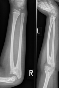
Fig 1.
Management
Indications for surgical treatment of congenital radioulnar synostosis are controversial. They are related to bilaterality and to the degree of deformity. Patients who have neutral rotation, mild pronation, or rare supination positions can compensate with ipsilateral shoulder motion. Wrist hypermobility can allow further functional compensation. Severe pronation deformities especially those more than 60º cause significant functional difficulty, especially with activities requiring supination. Hence, indications for surgery must be based more on individual functional limitations than on absolute forearm position.
Surgery should be performed in childhood before patients reach school age. In patients who have symptomatic subluxation of the radial head, the radial head may be excised at maturity.
The indication for surgery in posttraumatic radioulnar synostosis is forearm limitation of rotation. Surgery is performed after the synostosis has matured and distinct radiographic borders are seen. This will reduce the likelihood that the synostosis will recur. A too-long wait of more than 3 years will adversely affect the final outcome due to soft-tissue contracture. A 100º arc of motion is desirable so that the patient can perform all activities of daily living (ADLs), and a 60º arc is required to perform most ADLs without assistance.
The only contraindication for surgical correction is the presence of mild deformity in an older patient who has only minimal functional deficit.
Surgical Therapy
Attempts to achieve and maintain motion at the synostosis site are usually unsuccessful. Synostosis tends to recur despite excision, the use of various medications, or the interposition of fat, silicone, or muscle. Success, however, has been reported with excision of the bony bridge and the interposition of vascularized fat graft. An average rotation range of 74º can maintained at 2 years after surgery [24].
Some of the mobilization procedures are combined with tendon transfers to achieve supination. The extensor carpi radialis longus can be transferred to the volar aspect of the wrist and the flexor carpi ulnaris can be transferred dorsally around the ulna.
Congenital radioulnar synostosis
The preferred surgical procedure for the treatment of congenital radioulnar synostosis has been osteotomy and derotation through the fusion mass, along with fixation with transcutaneous pins [25,26,27].
The optimal position of correction will vary according to the degree of involvement present, the bilaterality of the synostosis, and the amount of compensatory shoulder or radiocarpal motion that the patient has. Severe deformities do not permit one-stage correction, because of the tension on fibrous and vascular structures. Gradual correction using a multiplanar external fixator decreases the risk of neurovascular injury and it allows the patient to select the most functional position.
Generally, neutral rotation is used for unilateral deformities, and for bilateral deformities, one side is placed in 20-30º of pronation and the other in 20-30º of supination [28]. Shortening of the forearm is recommended to decrease the risk of neurovascular compromise.
Postoperatively a long arm cast with 90º of elbow flexion is utilized for 8 weeks. After derotation, transcutaneous pins have generally been recommended for fixation. This pinning can easily be reversed if postoperative vascular compromise develops [8,29].
Simcock et al [30] reported the use of derotational osteotomy to treat 31 forearms in 26 children who had congenital radioulnar synostosis and functional limitations. In all cases, the union was successfully achieved within 8 weeks. There were no cases of compartment syndrome, vascular compromise, or loss of fixation. The overall rate of complications was 12%. This included two transient anterior interosseous nerve palsies in patients with rotational corrections of more than 80º, one transient radial nerve palsy, and one symptomatic muscle herniation.
Hwang et al [31] reported the use of one-stage rotational osteotomy of the proximal third of the ulna and the distal third of the radius with segmental bone resection to treat congenital radioulnar synostosis in 25 patients (28 forearms). There were 2 groups in this study. In group 1, the ulnar osteotomy was stabilized with an intramedullary pin. In group 2, no fixation was used. The surgical outcomes did not differ significantly between the two groups. The authors were of the opinion that one-stage rotational osteotomy of the proximal third of the ulna and the distal third of the radius with segmental bone resection was simple and safe and that internal fixation at the osteotomy site was not necessary.
Bishay [32] carried out a prospective study involving 12 consecutive pediatric patients (14 forearms) with severe congenital proximal radioulnar synostosis with a mean pronation deformity of 70.7° with a range of 60-85° that was corrected through single-session double-level rotational osteotomy and percutaneous placement of intramedullary Kirschner wires (K-wires) in both radius and ulna. At a mean follow-up of 30.4 months (range, 24-36 months), the patients had a mean pronation deformity correction of 59.8°. All 12 patients showed improvement in functional activities. None of the patients had any loss of correction or nonunion, neuropathies, circulatory disturbances, or hypertrophic scars.
Satake et al [33] carried out a study to assess the long-term (≥10 y) results of simple rotational osteotomy for congenital radioulnar synostosis in nine patients (12 forearms). Following the procedure, the forearm was fixed at an average of 4.2° of supination. At the final follow-up, the average motion arc of the palm ranged from 62° of supination to 26° of pronation. There were no postoperative neurologic or circulatory complications. The patients were better able to perform ADLs, and all were satisfied with the surgery results. The average score on the 11-item version of the Disability of the Arm, Shoulder, and Hand (DASH) score was 3.79 points at the final follow-up.
Pei et al [34] conducted a retrospective study of 31 patients (36 forearms) with congenital radioulnar synostosis. They found that proximal radioulnar derotational osteotomy followed by plate fixation was a safe and feasible procedure with a low complication rate. They were of the opinion that the technique could effectively improve the function of the forearm. Hamiti et al [35] also reported good results with this approach.
Posttraumatic radioulnar synostosis
The surgery for posttraumatic synostosis is different from that for congenital radioulnar synostosis. Surgery for posttraumatic synostosis restores motion through excision of the synostosis area [5,6,36]. Numerous interposition materials such as fat, fascia, silicone, muscle, and cellophane have been used after resection to prevent a recurrence of synostosis, but these have met with varying degrees of success. Kelikian and Doumanian [37] have developed a metallic swivel prosthesis to restore motion, but no large series has been reported that supports its effectiveness.
The goal of treatment involves resection of the entire bony synostosis.
The dissection should be carefully carried out with minimal periosteal disruption to prevent the further stimulation of bone, limiting recurrence. The protection and identification of neurovascular structures are essential. The final range of motion should be assessed intraoperatively. There should be minimal postoperative immobilization.
Giannicola et al [38] carried out a study to evaluate 12 patients with posttraumatic proximal radioulnar synostosis (two with a double synostosis) who were treated surgically. The synostosis was excised in 10 cases. Radial head excision was done in one case, radial head arthroplasty in three, and proximal radial diaphyseal resection in two. The mean extension-flexion and pronation-supination arcs were 116° and 123°, respectively. There were significant improvements in the Mayo Elbow Performance Score (mean, 24), modified American Shoulder and Elbow Surgeons score (mean, 28), and QuickDASH score (mean, 17). One patient had synostosis recurrence and one had late disassembly of the radial head arthroplasty.
Abdul Azeem et al [39] reported good functional and prophylaxis against recurrence results with a triple therapy consisting of preoperative radiation therapy, tissue interposition after heterotopic ossification resection, and postoperative adjuvant indomethacin.
Complications
Although the surgical procedure that is used to treat congenital radioulnar synostosis is quite straight forward, it is associated with significant complications [40,41]. These include neurovascular compromise and recurrence of ankylosis. Soft-tissue contracture and neurovascular compromise can occur. Simmons et al [22] were of the opinion that derotations of more than 85º should be performed in two stages. For suspected compartment syndromes a low threshold for fasciotomies should be maintained.
References
Rutkowski PT, Samora JB. Congenital Radioulnar Synostosis. J Am Acad Orthop Surg. 2021 Jul 1. 29 (13):563-570.
Wang E, Wenger DR, Zhang L, Zhao Q, Ji S, Li J. The mechanism of acute elbow flexion contracture in children with congenital proximal radioulnar synostosis. J Pediatr Orthop. 2010 Apr-May. 30 (3):277-81.
Shinohara T, Horii E, Tatebe M, Yamamoto M, Okui N, Hirata H. Painful snapping elbow in patients with congenital radioulnar synostosis: report of two cases. J Hand Surg Am. 2010 Aug. 35 (8):1336-9.
Sachar K, Akelman E, Ehrlich MG. Radioulnar synostosis. Hand Clin. 1994 Aug. 10 (3):399-404.
Hanel DP, Pfaeffle HJ, Ayalla A. Management of posttraumatic metadiaphyseal radioulnar synostosis. Hand Clin. 2007 May. 23 (2):227-34, vi-vii.
Watson FM Jr, Eaton RG. Post-traumatic radio-ulnar synostosis. J Trauma. 1978 Jun. 18 (6):467-8.
Mital MA. Congenital radioulnar synostosis and congenital dislocation of the radial head. Orthop Clin North Am. 1976 Apr. 7 (2):375-83.
Dawson HG. A congenital deformity of the forearm and its operative treatment. Br Med J. 1912. 2:833-5.
Hansen OH, Andersen NO. Congenital radio-ulnar synostosis. Report of 37 cases. Acta Orthop Scand. 1970. 41 (3):225-30.
Kelikian H. Congenital Deformities of the Hand and Forearm. Philadelphia: WB Saunders; 1974.
Lewis WH. The development of the arm in man. Am J Anat. 1901. 1:145-83.
Miura T, Nakamura R, Suzuki M, Kanie J. Congenital radio-ulnar synostosis. J Hand Surg Br. 1984 Jun. 9 (2):153-5.
Spritz RA. Familial radioulnar synostosis. J Med Genet. 1978 Apr. 15 (2):160-2.
Bauer G, Arand M, Mutschler W. Post-traumatic radioulnar synostosis after forearm fracture osteosynthesis. Arch Orthop Trauma Surg. 1991. 110 (3):142-5.
Henket M, van Duijn PJ, Doornberg JN, Ring D, Jupiter JB. A comparison of proximal radioulnar synostosis excision after trauma and distal biceps reattachment. J Shoulder Elbow Surg. 2007 Sep-Oct. 16 (5):626-30.
Garland DE, Dowling V. Forearm fractures in the head-injured adult. Clin Orthop Relat Res. 1983 Jun. (176):190-6.
Sauder DJ, Athwal GS. Management of isolated ulnar shaft fractures. Hand Clin. 2007 May. 23 (2):179-84, vi.
Cleary JE, Omer GE Jr. Congenital proximal radio-ulnar synostosis. Natural history and functional assessment. J Bone Joint Surg Am. 1985 Apr. 67 (4):539-45.
Jaffer Z, Nelson M, Beighton P. Bone fusion in the foetal alcohol syndrome. J Bone Joint Surg Br. 1981. 63B (4):569-71.
Giuffrè L, Corsello G, Giuffrè M, Piccione M, Albanese A. New syndrome: autosomal dominant microcephaly and radio-ulnar synostosis. Am J Med Genet. 1994 Jul 1. 51 (3):266-9.
Wilkie DP. Congenital radio-ulnar synostosis. Br J Surg. 1914. 1:366-75.
Simmons BP, Southmayd WW, Riseborough EJ. Congenital radioulnar synostosis. J Hand Surg Am. 1983 Nov. 8 (6):829-38.
Liu L, Liu C, Rong YB, Bai F, Chen SL. Radial Pronation Angle: A Novel Radiological Evaluation Index of Congenital Proximal Radioulnar Synostosis. Ann Plast Surg. 2020 May. 84 (5S Suppl 3):S196-S201.
Sonderegger J, Gidwani S, Ross M. Preventing recurrence of radioulnar synostosis with pedicled adipofascial flaps. J Hand Surg Eur Vol. 2012 Mar. 37 (3):244-50.
Ogino T, Hikino K. Congenital radio-ulnar synostosis: compensatory rotation around the wrist and rotation osteotomy. J Hand Surg Br. 1987 Jun. 12 (2):173-8.
Khalil I, Vizkelety T. Osteotomy of the synostosis mass for the treatment of congenital radio-ulnar synostosis. Arch Orthop Trauma Surg. 1993. 113 (1):20-2.
El-Adl W. Two-stage double-level rotational osteotomy in the treatment of congenital radioulnar synostosis. Acta Orthop Belg. 2007 Dec. 73 (6):704-9.
Green WT, Mital MA. Congenital radio-ulnar synostosis: surgical treatment. J Bone Joint Surg Am. 1979 Jul. 61 (5):738-43.
Smith RJ, Lipke RW. Treatment of congenital deformities of the hand and forearm (second of two parts). N Engl J Med. 1979 Feb 22. 300 (8):402-7.
Simcock X, Shah AS, Waters PM, Bae DS. Safety and Efficacy of Derotational Osteotomy for Congenital Radioulnar Synostosis. J Pediatr Orthop. 2015 Dec. 35 (8):838-43.
Hwang JH, Kim HW, Lee DH, Chung JH, Park H. One-stage rotational osteotomy for congenital radioulnar synostosis. J Hand Surg Eur Vol. 2015 Oct. 40 (8):855-61.
Bishay SN. Minimally invasive single-session double-level rotational osteotomy of the forearm bones to correct fixed pronation deformity in congenital proximal radioulnar synostosis. J Child Orthop. 2016 Aug. 10 (4):295-300.
Satake H, Kanauchi Y, Kashiwa H, Ishigaki D, Takahara M, Takagi M. Long-term results after simple rotational osteotomy of the radius shaft for congenital radioulnar synostosis. J Shoulder Elbow Surg. 2018 Aug. 27 (8):1373-1379.
Pei X, Han J. Efficacy and feasibility of proximal radioulnar derotational osteotomy and internal fixation for the treatment of congenital radioulnar synostosis. J Orthop Surg Res. 2019 Mar 20. 14 (1):81.
Hamiti Y, Yushan M, Yalikun A, Lu C, Yusufu A. Derotational Osteotomy and Plate Fixation of the Radius and Ulna for the Treatment of Congenital Proximal Radioulnar Synostosis. Front Surg. 2022. 9:888916.
Failla JM, Amadio PC, Morrey BF. Post-traumatic proximal radio-ulnar synostosis. Results of surgical treatment. J Bone Joint Surg Am. 1989 Sep. 71 (8):1208-13.
KELIKIAN H, DOUMANIAN A. Swivel for proximal radio-ulnar synostosis. J Bone Joint Surg Am. 1957 Jul. 39-A (4):945-52.
Giannicola G, Spinello P, Villani C, Cinotti G. Post-traumatic proximal radioulnar synostosis: results of surgical treatment and review of the literature. J Shoulder Elbow Surg. 2020 Feb. 29 (2):329-339.
Abdul Azeem M, Al-Hojailan K, Awad M, Khaja AF. Posttraumatic Radioulnar Synostosis: A Retrospective Case Series of 10 patients in Kuwait. J Shoulder Elbow Surg. 2022 Mar 9.
Barik S, Farr S, Gallone G, Zarantonello P, Trisolino G, Di Gennaro GL. Results after treatment of congenital radioulnar synostosis: a systematic review and pooled data analysis. J Pediatr Orthop B. 2021 Nov 1. 30 (6):593-600.
Hankin FM, Smith PA, Kling TF Jr, Louis DS. Ulnar nerve palsy following rotational osteotomy of congenital radioulnar synostosis. J Pediatr Orthop. 1987 Jan-Feb. 7 (1):103-6.