Thursday 28 September 2023

   Olecranon fractures


                                   Dr K Dhillon


Introduction

Ten percent of fractures around the elbow are olecranon fractures [1]. There is a diverse array of treatment options that have continued to evolve for the management of this fracture. These fractures vary in their complexity from relatively straightforward transverse fractures to comminuted and unstable configurations. No particular mode of treatment can be universally applied to the diverse array of fracture patterns encountered. For appropriate management of these fractures, the surgeon needs to have a good understanding of the anatomy, treatment options available, and potential complications.


Anatomy

The elbow is a complex hinge joint with a flexion arc of 0–150 degrees (fig1). The joint is stabilized by a number of factors. These include the anterior coronoid process and the posterior olecranon process which resist the translational forces of the humerus on the ulna.

The ulna collateral ligament and the radial head provide resistance to valgus stress. The lateral collateral ligament complex counters the varus stress.

Hyaline cartilage lines the articular surfaces. A transverse bare area devoid of cartilage is found at the midpoint between the coronoid and the tip of the olecranon [2]. The trochlea notch of the ulna, which articulates with the trochlea of the humerus has a transverse ‘‘bare area’’ at the junction between the anterior third and the posterior two-thirds.

This area varies in size between individuals. In one cadaveric study of 39 elbows, the mean width was 5.3 mm [3]. Knowledge of this area is important when reducing olecranon fractures. It can be tempting, but not correct, to eliminate any articular surface that is not covered by cartilage.

The triceps muscle inserts into the posterior third of the olecranon and the proximal ulna. It blends with the aponeurosis overlying the anconeus and the common extensor mechanism. The brachialis inserts into the coronoid process of the ulna and along with the triceps helps to produce compressive forces across the elbow joint during contraction.
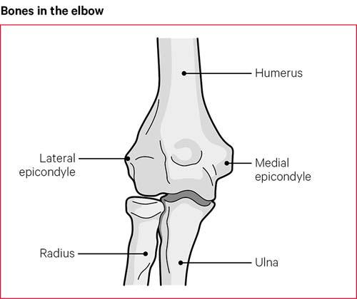

Fig 1.


Patella cubiti, an accessory ossicle embedded in the distal triceps may be present and can sometimes be mistaken for a fracture [4].

The ulnar nerve passes around the posterior aspect of the medial epicondyle and between the two heads of the flexor carpi ulnaris. The flexor carpi ulnaris spans in an arcade-like manner from the medial epicondyle to the olecranon process to form the roof of the cubital tunnel. The nerve lies posterior to the ulnar collateral ligament, which forms the floor of the cubital tunnel. The ulna collateral ligament attaches in a fanlike fashion to the medial border of the olecranon. The brachialis tendon inserts onto the proximal ulnar metaphysis distal to the tip of the midportion of the anterior coronoid.


Mechanism of injury

Direct or indirect trauma can cause fractures of the olecranon. A fall or blunt trauma on the posterior aspect of the elbow can cause a fracture directly. Indirect trauma from forces generated within the triceps muscle can occur with eccentric contraction during a fall on a partially flexed elbow leading to a fracture of the olecranon. Amis and Miller [5], in a cadaveric model, investigated the effect of impact mechanisms on olecranon fracture patterns. They found that the radial head and coronoid fractures occurred from impact to the forearm with the elbow in 80 degrees of flexion or less. Olecranon fractures followed direct blows at 90 of flexion. Distal humerus fractures were caused by impact when the elbow was in greater than 110 degrees of flexion. In cases of severe force to the elbow, a fracture dislocation can occur with posterior displacement of the olecranon fragment and the distal ulnar fragment together with the head of the radius. 


Classification

There are multiple classification systems for olecranon fractures. None have, however,  gained widespread acceptance. Colton et al [6] were the first, to develop a system based on the displacement and character of the fracture:

  • Type I fractures are undisplaced fractures

  • Type II fractures are unstable fractures. They are subdivided according to fracture pattern: type IIA avulsion fractures, type IIB transverse or oblique fractures, type IIC isolated comminuted fractures and type IID are fracture-dislocations. 

The AO classification of proximal radius and ulna fractures are divided into three broad groups. Type A are extra-articular fractures of either radius or ulna. Type B are intraarticular fractures of either bone, with type B1 being specifically an intra-articular fracture of the olecranon alone, and type C fractures are intra-articular fractures of both radius and ulna [7].

The Schatzker classification is based on the fracture pattern and a

consideration of the type of internal fixation that is required [8]. In type A the fracture line is transverse. In type B the fracture line is also transverse but there is impaction of bone. In type C the fracture line is oblique. In type D there is a comminuted fracture. In type E the fracture line is oblique and distal to the articular surface. In type F there is a fracture dislocation. 

The Mayo Clinic classification is one of the most frequently used and describes fractures on the basis of stability, displacement, and comminution:

  • Type I- the fractures are undisplaced and stable

  •  Type II- the fractures are displaced and unstable with intact collateral ligaments preventing dislocation 

  • Type III- the fracture is with an unstable elbow joint 

Type II and III fractures are further subdivided into A where the fracture is non-comminuted and B where the fracture is comminuted [9]. The Schatzker and Mayo classifications can be useful in predicting prognosis.  Schatzker types C (oblique) and D (comminuted) and Mayo type III fractures are associated with less favorable outcomes [10].


Diagnosis

History

Patients who have olecranon fractures and associated injuries present with pain and swelling around the elbow and distal arm. Patients with displaced fractures have an obvious deformity, and attempted motion can elicit painful crepitus. The patient is asked how the injury occurred. The patient is also asked about any associated neurovascular complications. A history of any concurrent illnesses precipitating the injury is obtained. A detailed account of comorbid conditions is important.

Clinical examination

An assessment of the soft tissues around the elbow is carried out during physical examination. There is usually extensive swelling with ecchymosis. The presence of abrasions or lacerations should be noted. There should be no aggressive assessment of range of motion or strength of the elbow.

A palpable sulcus may be present at the site of the olecranon fracture. There will be painful limitation of the range of motion of the elbow. There will be an inability to extend the elbow actively against gravity. A careful neurovascular examination is carried out before any planned manipulation of the elbow.

Radiographic and imaging assessment

Plain radiographs usually provide sufficient information for an accurate diagnosis. Severe comminution with displacement and overlap of bone fragments can obscure the fracture pattern. Radiographs have to be of good quality, out of splint and obtained while maintaining gentle longitudinal traction with the inclusion of the elbow joint on the film. Poor  X-rays done in a splint are not as well suited for accurate diagnosis, classifying the fracture, and formal preoperative planning. Radiographs are carefully evaluated for the presence of associated injuries, such as a radial head fracture or dislocation, a distal humerus fracture, or a coronoid fracture. A CT scan rarely provides additional information that helps in decision making, and preoperative planning with an isolated olecranon fracture. It should be reserved for more complex fractures. 


Treatment

Nondisplaced fractures of the olecranon (Mayo type IA and IB) are treated conservatively. Nondisplaced fractures are those where the displacement is less than 2 mm and there is no change in position with gentle flexion to 90 or extension of the elbow against gravity. These fractures are treated with a

long arm cast with the elbow in 90 of flexion for 3 to 4 weeks. After the cast is removed protected range of motion exercises are started. Flexion past 90 degrees is avoided until bone healing is complete at approximately 6 to 8 weeks. In elderly patients, the range of motion exercises can be started earlier than 3 weeks if patients can tolerate it so that stiffness can be prevented. After application of the cast, a follow-up radiograph should be done within 5 to 7 days to ensure that displacement of the fracture has not occurred. Immobilization in full extension is not done because stiffness is more likely. Fractures that require full extension for reduction should be treated surgically.

Tension band wire

Displaced olecranon fractures require surgical treatment to restore joint

congruity, elbow extension, and elbow stability. Transverse fractures without comminution (Mayo type IIA) can be treated by tension band wiring. A tension band wire construct converts the tensile distraction force of the triceps into a dynamic compressive force across the olecranon articular surface. K-wires are used for tension band wiring. Intramedullary cancellous screw fixation cannot be used in elderly patients who have osteoporosis. 

Two 1.6-mm or 2.0-mm K-wires are inserted into the olecranon tip and the wires engage distally in the anterior cortex of the ulna [11]. Overpenetration of the wires should be avoided to prevent neurovascular damage, limitation of forearm rotation, or heterotopic ossification. Once the wire penetrates the far cortex, it is partially backed out and bent 180 degrees at the previously noted position and cut. The fibers of the triceps tendon are split sharply with a scalpel at the site of the K-wires to allow the cut and bent ends to be impacted against the cortex. A figure-of-8 loop of 18-gauge or 1.5-mm gauge wire is passed through a drill hole located distally. The hole is drilled approximately and equal distance from the fracture as the tip of the olecranon. The wire is then passed deep to the fibers of the triceps, near the bone, beneath the K-wires. The wire is then tightened by twisting in two places on opposite arms of the crossed portion of the figure-of-8.

The K-wires are seated firmly in the bone using an impactor, beneath the fibers of the triceps. 

In transverse fractures with comminution (Mayo type IIB), the tension band technique cannot be used. It will collapse the fragments together, leading to a narrowed olecranon articulation that does not track properly. These fractures are treated with limited-contract dynamic compression (LCDC) plate fixation, with or without bone graft [12]. Similarly, a plate with lag screw fixation is preferred for oblique fractures or unstable displaced olecranon fracture–dislocations with and without comminution (Mayo type IIIA and IIIB). 


Limited-contract dynamic compression plate fixation

LCDC plate fixation has several advantages [13,14]. The plate allows improved contouring. It can be appropriately placed on the dorsal tension surface of the proximal ulna around the tip of the olecranon. It helps to hold the proximal fragment when poor bone quality limits screw purchase. The redesigned screw holes allow greater angulation of screw placement and the option of compression from either side of the screw hole. Furthermore, its lower profile allows its use in subcutaneous situations where soft tissue coverage is minimal. Fixation of the plate proximally is often the greatest challenge because the bone there can be thin, and cancellous screws rather than cortical screws are used in elderly patients. The newer precontoured plates allow for an increased number of fixation points in the proximal fragment. In complex fractures of the proximal ulna, a large coronoid fragment is usually present. This fragment is very important to the final stability of the elbow. It must be fixed with lag screws placed either through or adjacent to the implanted plate [15]. Mobilizing the proximal olecranon fragment allows the coronoid to be visualized and reduced through the olecranon fracture.

Technique

The patient is placed in a lateral position on the operating table with the injured arm on a bolster across the chest. A tourniquet is placed on the upper arm. Skin preparation and draping is carried out. A posterior midline incision centered on the olecranon is made and extended proximally 5 cm from the tip of the olecranon. The ulna is exposed along its subcutaneous border. The anconeus can be elevated to approach the radial head if required. Impacted articular fragments are elevated. The coronoid is then reduced and provisionally fixed to the ulna with K-wires. A narrow 3.5-mm LCDC plate is then contoured to fit the proximal ulna. The plate is bent near 90, between the second and third screw holes of the plate. After the fracture is reduced the contoured plate is applied to the dorsal aspect of the olecranon. The plate is fixed proximally with one screw obliquely upward into the coronoid process. Fixation of the coronoid can also be performed with lag screws adjacent to the plate. Additional screws are placed proximally in the olecranon. The plate is secured distally to the shaft with three or four bicortical screws.

In patients with osteoporotic olecranon, direct trauma to the posterior aspect of the elbow can lead to severely comminuted fractures. In such situations, excision of the fracture fragments and reattachment of the triceps tendon may be necessary in elderly patients whose olecranon fracture fragments are too small or too comminuted for internal fixation.

The coronoid and anterior soft tissues, collateral ligaments, and interosseus membrane must be intact. The triceps tendon is reattached adjacent to the articular surface with nonabsorbable sutures that are passed through drill holes in the proximal ulna. Reattaching the triceps this way creates a sling for the trochlea. There is a smooth congruent transition from the triceps tendon to the articular surface but it decreases the moment arm. This can result in a weaker extensor mechanism but elbow stability is enhanced [16]. The amount of olecranon that can be safely excised remains debatable. Based on in vitro [17] and clinical studies [18,19] between 50% and 70% of the olecranon articular surface can be excised without compromising elbow stability as long as the coronoid and distal trochlea are preserved.


Complications

Painful hardware irritation is one of the most common complication after

internal fixation of olecranon fractures. Such a complication has been reported in up to 80% of cases. The incidence of prominent painful hardware is more after tension band wiring as compared to compression plating [20,21]. Simpson and colleagues [13] reported no cases of symptomatic hardware irritation after LCDC plating. Bailey and colleagues [22], however, reported that 20% of their patients required plate removal because of the prominence of the plate fixation.

Loss of movements is usually not a significant problem in patients with isolated olecranon fractures. In patients with isolated olecranon fractures, the typical loss of motion is 10 to 15 degrees of extension. Patients who have associated fractures of the radial head or coronoid are more likely to develop limitations of motion.

Nonunion of olecranon fractures has been reported in up to 1% of the patients. The typical symptoms of nonunion are pain, instability, or loss of motion [23]. Treatment options for olecranon nonunions

include:

  • Excision of the olecranon fragment.

  • Osteosynthesis with a LCDC plate, and bone grafting.

  • Elbow arthroplasty in the presence of severe posttraumatic arthritis. 


In elderly patients, acceptable management includes excision of the proximal portion of the pseudarthrosis and repair of the triceps tendon, making sure that the coronoid and anterior soft tissues are intact.


Rehabilitation

Early initiation of physical therapy is one of the most important issues in

elbow surgery especially in the elderly. Postoperatively, a posterior POP slab with the elbow flexed to 90 degrees is applied to help manage postoperative pain. The posterior slab is usually discontinued after 5 to 7 days and a removable splint is provided to the patient. Gentle active assisted exercises and passive motion exercises are then started. The patient is told to support the wrist with the opposite hand and gently flex and extend the elbow. The arm is taken out of the splint several times a day for these exercises and to let gravity work on extending the elbow. Active motion against resistance is avoided until callus formation is evident. This usually happens at 8 to 10 weeks. If the stability of the fixation is in doubt, then a hinged fracture brace can be used to provide additional support.


Results and outcome

The clinical outcomes after fractures of the olecranon are generally good to excellent. Most series report satisfactory outcomes and restoration of normal or near-normal function in more than 95% of patients.

Bailey et al [22] evaluated the functional outcome of plate fixation for displaced olecranon fractures (Mayo type II or III) in 25 patients at an average follow-up of 34 months. They found that the patient satisfaction was high (9.7/10) with a low pain rating (1/10). Based on the Mayo elbow score, 22 patients had excellent or good outcomes. The mean DASH score showed almost normal upper extremity function.

Karlsson et al [24] evaluated the long-term outcome of closed olecranon fractures in 73 patients at a mean of 19 years after the fracture. The primary treatment consisted of open reduction and internal fixation in 84% of the elbows. Of these patients, 61 had no complaints at follow-up, 9 had occasional pain, and 3 had daily pain. Ninety-six percent of the patients had an excellent or good overall outcome.


Conclusion

About 10% of the fractures around the elbow are olecranon fractures. There is a diverse array of treatment options that have continued to evolve for the management of this fracture. Some of these fractures are relatively straightforward transverse fractures and others are comminuted and unstable. No particular mode of treatment can be universally applied to the diverse array of fracture patterns encountered. For appropriate management of these fractures, the surgeon needs to have a good understanding of the anatomy, treatment options available, and potential complications.

Olecranon fractures have good to excellent outcomes with adherence to a treatment algorithm based on displacement, comminution, and joint stability. Decreased range of motion, radiographic evidence of degenerative changes, and requirement for hardware removal are common. These can be minimized through careful attention to proper technique, anatomic reduction with stable fixation, and early mobilization.


References

  1. Rommens PM, Kuchle R, Schneider RU, Reuter MM. Olecranon fractures in adults: factors influencing outcome. Injury 2004; 35:1149–57.

  2. Stormont TJ, An KN, Morrey BF, et al. Elbow joint contact study: comparison of techniques. J Biomech 1985;18(5):329–36.

  3. Wang A, Mara M, Hutchinson DT. The proximal ulna: an anatomic study with relevance to olecranon osteotomy and fracture fixation. J Shoulder Elbow Surg 2003;12(3):293–6.

  4. Winter M, Balaguer T, Tabutin J. Bilateral patella cubiti. A case report. J Bone Joint Surg Am 2006; 88(2):415–7.

  5. Amis AA, Miller JH. The mechanisms of elbow fractures: an investigation using impact tests in vitro. Injury 1995;26(3):163–8.

  6. Colton CL. Fractures of the olecranon in adults: classification and management. Injury 1973;5(2):121–9.

  7. Lavigne G, Baratz M. Fractures of the olecranon. J Am Soc Surg Hand 2004;4(2):94–102.

  8. Browner BD, Jupiter JB, Levine AM, et al. Skeletal trauma. Philadelphia: Saunders; 1992.

  9. Morrey BF. Current concepts in the treatment of fractures of the radial head, the olecranon, and the coronoid. J Bone Joint Surg Am 1995; 77:316–27.

  10. Rommens PM, Kuchle R, Schneider RU, Reuter MM. Olecranon fractures in adults: factors influencing outcome. Injury 2004; 35:1149–57.

  11. Prayson MJ, Williams JL, Marshall MP, et al. Biomechanical comparison of fixation methods in transverse olecranon fractures: a cadaveric study. J Orthop Trauma 1997;11(8):565–72.

  12. Hak DJ, Golladay GJ. Olecranon fractures: treatment options. J Am Acad Orthop Surg 2000;8(4): 266–75.

  13. Simpson NS, Goodman LA, Jupiter JB. Contoured LCDC plating of the proximal ulna. Injury 1996; 27(6):411–7.

  14. McKee MD, Seiler JG, Jupiter JB. The application of the limited contact dynamic compression plate in the upper extremity: an analysis of 114 consecutive cases. Injury 1995;26(10):661–6.

  15. Doornberg J, Ring D, Jupiter JB. Effective treatment of fracture-dislocations of the olecranon requires a stable trochlear notch. Clin Orthop Relat Res 2004;429:292–300.

  16. Coonrad RW, Morrey BF. Management of olecranon fractures and nonunion. In: Morrey BF, editor. Master techniques in orthopaedic surgery: the elbow. 2nd edition. Philadelphia: Lippincott Williams & Wilkins; 2002. p. 103–26.

  17. An KN, Morrey BF, Chao EY. The effect of partial removal of proximal ulna on elbow constraint. Clin Orthop Relat Res 1986;209:270–9.

  18. Inhofe PD, Howard TC. The treatment of olecranon fractures by excision or fragments and repair of the extensor mechanism: historical review and report of 12 fractures. Orthopedics 1993;16(12): 1313–7.

  19. Gartsman GM, Sculco TP, Otis JC. Operative treatment of olecranon fractures. Excision or open reduction with internal fixation. J Bone Joint Surg Am 1981;63(5):718–21.

  20. Hume MC, Wiss DA. Olecranon fractures. A clinical and radiographic comparison of tension band wiring and plate fixation. Clin Orthop Relat Res 1992;285:229–35.

  21. Wolfgang G, Burke F, Bush D, et al. Surgical treatment of displaced olecranon fractures by tension band wiring technique. Clin Orthop Relat Res 1987;224:192–204.

  22. Bailey CS, MacDermid J, Patterson SD, et al. Outcome of plate fixation of olecranon fractures. J Orthop Trauma 2001;15(8):542–8.

  23. Papagelopoulos PJ, Morrey BF. Treatment of nonunion of olecranon fractures. J Bone Joint Surg Br 1994;76(4):627–35.

  24. Karlsson MK, Hasserius R, Karlsson C, et al. Fractures of the olecranon: a 15- to 25-year followup of 73 patients. Clin Orthop Relat Res 2002;403:205–12.

Tuesday 19 September 2023

   Reflex Sympathetic Dystrophy


                              DR KS Dhillon


Introduction

Complex Regional Pain Syndrome (CRPS) is a neuropathic pain disorder. It is characterized by persistent pain that is disproportionate to the degree of tissue injury and persists beyond the usual expected time for tissue healing [1].  Pain is accompanied by motor, sensory, and autonomic abnormalities. These abnormalities include hyperalgesia, allodynia, sudomotor, and vasomotor abnormalities, as well as trophic changes. The pain does not follow a particular dermatome or myotome but is rather regional. This disabling condition usually develops after a fracture, trauma, or surgery [2][3]. Some spontaneous cases have also been reported [4].

Ambroise Paré in the 16th century, reported cases with CRPS-like symptoms for the first time which developed following phlebotomy [5]. Silas Mitchell in 1864, observed this syndrome after gunshot wounds. In 1864 he used the term ‘causalgia’ to describe this syndrome. In 1946 James A. Evans coined the term ‘reflex sympathetic dystrophy’ to describe a similar condition. He suspected that the pain was sympathetically mediated [6]. In 1994, the International Association for the Study of Pain (IASP) named this condition ‘Complex Regional Pain Syndrome’. The IASP proposed a diagnostic criterion. Due to low specificity, a widely accepted revised criterion was proposed in 2010. It is commonly referred to as the "Budapest Criteria" [2,7].

CRPS has two subtypes. Type I was formerly known as reflex sympathetic dystrophy, and type II was formerly known as causalgia. In type I there is no nerve trauma. Type II occurs in the setting of known nerve trauma. Clinically they are indistinguishable. They follow a regional rather than a dermatomal or peripheral nerve distribution and favor the distal extremities. Spread outside of the initially affected area commonly occurs to the proximal or contralateral limb [6,8]. CRPS is further subdivided into "cold" versus "warm," and sympathetically maintained (SMP) versus sympathetically-independent (SIP), which may affect prognosis and treatment options [8].

CRPS not only affects sleep, function, and activities of daily living but also takes a significant mental and psychosocial toll on the patient [9,10,11]. Its diverse spectrum of clinical presentation and lack of clearly defined pathophysiology poses a challenge for optimal management. 


Etiology

CRPS usually occurs due to varying degrees or types of tissue trauma. It has been documented even in the absence of injury. It has been seen following periods of prolonged immobilization. A fracture is the most common injury associated with developing CRPS. Surgery is another common cause. Other inciting injuries or insults include contusions, sprains, and crush injuries. CRPS has even been reported after seemingly innocuous interventions such as intravenous line placement. Increased psychological distress experienced during the physical injury may affect the severity and prognosis of CRPS. 


Fracture

CRPS is commonly associated with extremity fractures. A large multicenter prospective study by Beerthuizen et al [12] found that 48.5% of patients developed CRPS (IASP criteria) after suffering a single fracture of the ankle, wrist, scaphoid, or the fifth metatarsal. All the patients remained symptomatic at 1-year follow-up. They found that rheumatoid arthritis and intraarticular ankle fractures and dislocations were risk factors for CRPS. There was no significant difference for disease onset between fractures of arms or legs.

Another prospective cohort study by Brunner et al [13] found that CRPS developed within 8 weeks after a noxious event. Symptoms improved in many patients at 3 months. There was no significant improvement noted at a year.

There are studies involving patients who developed CRPS after fracture of the distal radius that have identified higher age, social or psychological factors, and psychiatric comorbidities as risk factors [14,15].

There was, however, another prospective study that did not find any correlation between psychological factors or depression and the development of CRPS [16].


Surgery

Extremity surgeries are also more commonly associated with the development of CRPS. In a retrospective study by Rewhorn et al [17] of 390 patients who underwent foot and/or ankle surgeries, 4.36% developed CRPS. Surgical treatment of fractures has been found to have a higher risk of CRPS. 

A study by JelladIn et al [18] found that 32.2 % of their patients undergoing closed reduction of distal radius fracture, developed CRPS. Carpal tunnel surgeries were noted to have a 2 to 5% incidence of CRPS and Dupuytren contracture surgeries had a 4.5 to 40% incidence of CRPS [19]. 


Genetics

The impact of genetic factors in the development of CRPS remains unclear. Tumor necrosis factor-alpha (TNF-α) polymorphism and human leukocyte antigen have been found to play a role in CRPS. When these factors are present there can be an earlier age of onset and more severe symptoms. A few retrospective reports have suggested familial inheritance [19].


Epidemiology

The incidence of CRPS appears to vary based on geographical location. A study by Sandroni et al. in Olmsted County Minnesota, that was reported in 2003, found an incidence of 5.46 per 100,000 person-years for CRPS type I and 0.82 per 100,000 person-years for CRPS type II [20]. Another study by Mos et al. in the Netherlands reported in 2006, found the incidence to be much higher at 26.2 cases per 100,000 person-years [21]. Both studies found that females were more often affected. The first study found that females were four times more likely to be affected than males, while the second study found that this disorder was at least three times more common in females [20,21].


The Netherlands study reported a peak incidence at 61–70 years of age. The American study found the median age of onset to be 46 years. Upper limbs were more commonly involved than lower limbs in both studies. Both studies used the IASP CRPS criteria for diagnosis of the disease. The most common trigger for the disease was found to be a fracture. A fracture was associated with 44 to 46% of the cases. Vasomotor symptoms of swelling, temperature, and color changes were most commonly reported [20,21].

The three-phase bone scans are most useful for making a diagnosis (85%). Autonomic testing is helpful in making a diagnosis in 80% of cases [20]. Asthma, menopause, angiotensin-converting enzyme (ACE) inhibitor use, osteoporosis, and history of migraine are risk factors for CRPS [21,22]. Cigarette smoking also increases the risk of developing CRPS [23].


Pathophysiology

Multiple pathophysiologic mechanisms have been described to explain CRPS. Scientific evidence does not point to a single main mechanism. The underlying mechanism seems to be multifactorial. Immunological, inflammatory, central, and peripheral sensitization, as well as autonomic changes, have been studied in CRPS [6].

Inflammatory Changes

Both the clinical presentation as well as the elevated inflammatory markers suggest that inflammation is a key mechanism underlying the development of CRPS. The basic features of inflammation, such as swelling, redness, increased temperature, pain, and functional impairment, are commonly associated with CRPS [24]. Elevated levels of pro-inflammatory cytokines such as TNF-α, Interleukin (IL)-1b, IL-2, and IL-6 have been found in both serum and cerebrospinal fluid of patients with CRPS [25,26,27,28]. As a result of tissue injury elevated levels of neuropeptides like calcitonin gene-related peptide (CGRP), bradykinin, and substance P are released from peripheral nerve endings. They trigger neurogenic inflammation. The elevated levels of inflammatory markers and neuropeptides cause vasodilation and tissue extravasation [22,29,30,31,32].

Immunological Changes

Autoimmune factors play a role in CRPS pathogenesis. Autoantibodies against beta-2-adrenergic receptors, muscarinic-2 receptors, and alpha -1a-adrenergic receptors, have been found in CRPS [33,34]. Goebel et al [35] found a significant improvement in pain following intravenous immunoglobulin treatment in CRPS patients. This further supports potential autoimmune pathophysiology.

Peripheral Sensitization

Peripheral nervous system sensitization is triggered by the release of pro-inflammatory markers after the injury. Markers such as TNF-α released reduce the stimulation threshold. This leads to local sensitization and hyperalgesia in CRPS. Catecholamine sensitivity of peripheral nerve fibers has also been seen in CRPS [6].

Central Sensitization and Neuroplasticity

In patients with CRPS, increased excitability of secondary dorsal horn neurons occurs. As a result of sensitization hyperalgesia and allodynia develops. The release of bradykinin, substance-P, and glutamate plays an important role in this process. Continued noxious primary afferent traffic into the dorsal horn leads to wind-up and central sensitization [24]. Based on the response to ketamine infusions in CRPS patients, activation of spinal N-methyl D-aspartate (NMDA) receptors seems to play an important role in the pathogenesis [36,37]. Improvement of CRPS symptoms with intrathecal baclofen suggests gamma-aminobutyric acid (GABA) involvement in sensitization [6].

Evidence of cortical reorganization has been noted in CRPS. A reduction in the somatosensory-cortex area corresponding to the affected extremity occurs [38]. The degree of neuroplasticity correlates with the intensity of pain and severity of hyperalgesia, both of which indicate central sensitization [39,40].

Autonomic Changes

Sympathetic-afferent coupling occurs in CRPS. It is due to the upregulation of sympathetic receptors on nociceptive nerve fibers. As a result of this sympathetic hyperactivity, there is increased pain and sympathetic sensitivity of nociceptive nerves. The local swelling, color changes, and temperature variations associated with this disorder suggest an involvement of the autonomic nervous system [41]. Widespread autonomic dysregulation in CRPS can affect the heart rate and can lead to orthostatic dysfunction [42]. In warm CRPS, vasodilation occurs as a result of reduced catecholamine release. The opposite phenomenon occurs in cold CRPS [6].


History and Physical Examination

Patients can have sensory, motor, or autonomic symptoms. Sensory symptoms include allodynia where non-painful stimuli cause pain and hyperalgesia where painful stimuli cause exaggerated pain. Patients can also have autonomic symptoms. These include skin color and temperature changes (vasomotor dysfunction) and swelling and sweating changes (sudomotor dysfunction). Motor symptoms include weakness, tremors, reduced range of motion, and even dystonia (involuntary muscle contraction) in the affected extremity [43].

CRPS can be associated with worsening depression, poor function, anxiety, and diminished quality of life. A systematic review by Lohnberg et al [11] examined psychosocial factors associated with CRPS and they concluded that there is no support in the literature for specific personality or psychopathology predictors of CRPS [11]. Patients with a significant comorbid psychological burden and/or poor coping mechanisms can demonstrate pain-related behavior and catastrophic thinking. 

CRPS can also be associated with systemic medical conditions such as neuropsychological deficits that include executive functioning, memory, word retrieval, constitutional symptoms such as lethargy, weakness, disruptions in sleep architecture, cardiopulmonary involvement which includes neurocardiogenic syncope, atypical chest pain, chest wall muscle dystonia leading to shortness of breath, endocrinopathies which include impaired hypothalamic-pituitary-adrenal axis with low serum cortisol, hypothyroidism, urologic dysfunction with increased urinary frequency and urgency, urinary incontinence, and gastrointestinal dysmotility with nausea, vomiting, diarrhea, constipation, indigestion [44,45,46,42,47,48].


Evaluation

The pathophysiologic mechanism for CRPS has yet to be identified. There is no gold standard diagnostic test for CRPS [8]. The diagnosis is clinical. It is based on the widely accepted Budapest criteria. As compared to the previous IASP criteria, the Budapest criteria have similar sensitivity (0.99) but higher specificity (0.68) [7].

The Budapest Criteria [6] includes the following:

A. There should be continuing pain that is disproportionate to the inciting event.

B. There should be at least one symptom in three of the following four categories:

  • Sensory: Reports of hyperalgesia and/or allodynia.

  • Vasomotor: Reports of temperature asymmetry and/or skin color changes and/or skin color asymmetry.

  • Sudomotor/edema: Reports of edema and/or sweating changes and/or sweating asymmetry.

  • Motor/trophic: Reports of decreased range of motion and/or motor dysfunction (weakness, tremor, dystonia) and/or trophic changes (hair, skin, nails).

C. They must display at least one sign at the time of evaluation in two or more of the following categories:

  • Sensory: Evidence of hyperalgesia (to pinprick) and/or allodynia (to light touch or deep somatic pressure),

  • Vasomotor: Evidence of temperature asymmetry and/or skin color changes and/or asymmetry,

  • Sudomotor/edema: Edema and/or sweating changes and/or sweating asymmetry,

  • Motor/trophic: Evidence of decreased range of motion and/or motor dysfunction (weakness, tremor, dystonia) and/or trophic changes (hair, skin, nails).

D. There is no other diagnosis that better explains the signs and symptoms.


Various objective testing measures such as thermography, triple-phase bone scan, and the quantitative sudomotor axon reflex test have been utilized. They are, however, not necessary to make the diagnosis of CRPS. The diagnosis of CRPS is largely clinical. The differential diagnosis of CRPS includes small or large fiber sensorimotor neuropathy, vasculitis, vascular insufficiency, lymphedema, cellulitis, erythromelalgia, deep vein thrombosis, and Reynaud’s phenomenon. Diagnostic tests in CRPS are done to screen for other potential differential diagnoses.


Management

Patients with CRPS can improve spontaneously. In some patients, the symptoms can be debilitating. It is good to institute aggressive management as soon as possible since a delay may result in an unfavorable outcome. Compared to chronic CRPS, early CRPS is less resistant to treatment and has a better prognosis [49]. The aim of treatment is not only to improve pain and discomfort but also to restore function and prevent disability. The most optimal treatment would include an interprofessional approach including physical and occupational therapy, pharmacotherapy, behavioral therapy, and interventions [6].


Physical and Occupational Therapy

Manual therapy and exercises are part of the treatment regime for CRPS. Other treatment modalities include ultrasound, laser, pain education, transcutaneous electrical nerve stimulation, mirror therapy, and graded motor imagery (GMI). Manual therapy and exercise improve the function, and range of motion, as well as reduce disability through endorphin release as well as other central and peripheral analgesic mechanisms [3,50]. Pain education influences pain perception and behavior by improving understanding of pain pathophysiology [3]. The GMI and mirror therapy remediate maladaptive cortical neuroplastic changes that are associated with chronic pain conditions like CRPS [51].

A 2016 Cochrane review found that GMI and mirror therapy may improve pain as well as function in CRPS. The quality of the evidence, however, was poor. Two clinical trials each for mirror therapy and GMI therapy have demonstrated improvement in pain and function at 6 months. Low-quality evidence was also found for the improvement of impairment in CRPS with multimodal physiotherapy [3].


Pharmacotherapy

There are several medications that are used in the management of CRPS. These include anti-inflammatory medications, antidepressants, transdermal lidocaine, anticonvulsants, opioids, bisphosphonates, and NMDA antagonists. Using a multimodal pharmacologic regimen can lead to superior outcomes. 

Anti-inflammatory Medications

Since inflammation is thought to play a role in disease pathogenesis, oral corticosteroids and non-steroidal anti-inflammatory drugs (NSAIDs) have been used in CRPS. There were three trials that compared oral corticosteroids to placebo in CRPS. Based on these trials a 2013 Cochrane review concluded that oral steroids do not significantly reduce pain. This, however, was supported by very low-quality evidence. The Cochrane review also found that oral corticosteroids do seem to improve composite pain scores [52]. Another study compared piroxicam (NSAID) to oral prednisone. The study found that oral prednisolone seemed to be more effective in improving composite CRPS scores in post-stroke patients [53]. A study by Kalitamore et al [54] found that a 2-month treatment with low-dose oral prednisone was safe and effective in post-stroke CRPS.

Bisphosphonates

Bisphosphonates such asrisedronate (Actonel), alendronate (Fosamax), ibandronate (Boniva), zoledronic acid (Reclast), and pamidronate (Aredia) are routinely used in bone-related problems as it inhibits osteoclastic activity. There are several mechanisms of action of bisphosphonates in CRPS. The more commonly accepted mechanism includes inhibition of bone marrow cell proliferation and migration. It also inhibits inflammation modulation [55]. A 2017 meta-analysis concluded that bisphosphonates reduce pain in CRPS I [56]. A Cochrane review in 2013 found that there is low-quality evidence that also seemed to suggest the same response in CRPS. It is more so in those with concomitant evidence of osteopenia or osteoporosis [52].

Anticonvulsants and Antidepressants

Gabapentin, an anticonvulsant, is the most widely studied medication in this class. Its mechanism is via inhibition of the alpha 2-delta subunit of voltage-gated calcium channels. It is widely used in the treatment of CRPS, although the quality of evidence regarding its effectiveness in treating CRPS is very low [52]. A study in 2016 compared amitriptyline and gabapentin for CRPS I and pediatric neuropathic pain. The study found that both the medications reduced pain intensity and disability significantly. However, there was no significant difference in effect between the two [57].

Opioids

The effectiveness of opioids for the treatment of CRPS has not been studied. Therefore no evidence-based conclusions can be drawn [41].

NMDA Antagonists

NMDA receptor antagonists such as ketamine have been hypothesized to reverse central sensitization and maladaptive cortical neuroplastic changes in patients with CRPS [24]. There is low-quality evidence which suggests that intravenous ketamine infusion may improve pain in CRPS for up to 4-11 weeks [52,52]. Side effects and psychomimetic properties of ketamine have prevented its widespread use [24].


Behavioral Therapy

In patients with depression, the levels of catecholamines are elevated and this can worsen CRPS by inducing central sensitization through adrenergic mechanisms. Psychotherapy can help reverse this effect. There is only one small trial that has evaluated the efficacy of behavioral interventions in CRPS. Despite the lack of clear evidence supporting the use of behavioral therapy in CRPS, behavioral therapy has been recommended as part of comprehensive treatment [58].


Interventions

Sympathetic Blocks

Sympathetic hyperactivity is believed to cause CRPS [41]. Therefore, lumbar sympathetic nerve blocks are used to treat lower extremity symptoms, and stellate ganglion sympathetic blocks are used to treat upper extremity symptoms of this syndrome. A 2013 Cochrane review found that sympathetic blocks with local anesthetic were ineffective at reducing CRPS related pain. The quality of evidence was, however, low [52]. Another Cochrane review in 2016 failed to draw any definitive conclusions on the efficacy of such treatment in CRPS due to paucity of evidence [59].


Spinal Cord Stimulation

Spinal cord stimulation (SCS) is carried out by delivering electric stimulation to the dorsal column of the spinal cord by the placement of electrodes in the epidural space. The electrodes are connected to an implanted pulse generator to power the electrode. In some devices, an external pulse generator is used.

Several mechanisms of action of SCS have been proposed. This includes inhibition of nociceptive neural conduction in the spinal cord, vasodilation, adrenergic inhibition, and reversal of cortical maladaptive neuroplastic changes. There was a systematic review in 2017 that studied the effectiveness of SCS in CRPS. The authors of the review concluded that a high level of evidence supports the use of SCS for the improvement of pain scores, quality of life as well as the perception of pain relief in CRPS [59].

Dorsal Root Ganglion Stimulation

Targeting the dorsal root ganglion (DRG) instead of the spinal cord is a new and novel neuromodulation modality for the treatment of chronic pain. This allows a more focused application of neurostimulation than traditional SCS. DRG stimulation was approved by the United States Food and Drug Administration in 2016 for the treatment of lower extremity pain in CRPS.

A pooled analysis study by Huygen et al [60] concluded that DRG stimulation was safe and effective for CRPS with a 4.9-point mean reduction of pain intensity in CRPS-I. The ACCURATE study compared SCS and DRG stimulation in 152 subjects with CRPS. The study results were published in 2017. This randomized trial found that DRG stimulation was more effective than traditional SCS in reducing pain and improving quality of life in CRPS [61].


Differential Diagnosis

The differential diagnosis of CRPS includes:

  • Arterial insufficiency

  • Gillian Barre syndrome

  • Hysteria

  • Phlebothrombosis

  • Porphyria

  • Poliomyelitis

  • Tabes dorsalis

  • Monometric amyotrophy

  • Multiple sclerosis

  • Peripheral atherosclerotic disease


Staging

In 1990 Bonica proposed 3 stages of CRPS. Bruehl et al [62] studied the validity of the 3 stages in a series of 113 patients and they found no significant difference in duration of symptoms among the stages. This suggests that clear generalized disease stages don't exist in CRPS.


Prognosis

The prognosis of CRPS is variable. Spontaneous remission and refractory clinical presentation have been seen in CRPS. However early treatment may improve the prognosis.


Complications

Some of the complications seen in patients with long-standing CRPS include:

  • Dystonia

  • Cognitive executive dysfunction

  • Irritable bowel syndrome 

  • Adrenal insufficiency

  • Gastroparesis


Deterrence 

It has been hypothesized that oral supplementation of vitamin C lowers the risk of the development of CRPS after fractures due to its antioxidant properties. In 2015, a meta-analysis of 3 trials found that the available evidence failed to demonstrate a definitive preventive role of vitamin C in CRPS development after distal radial fractures. The level of evidence was, however, low [63]. In 2017 another meta-analysis and systemic review evaluated the efficacy of vitamin C in the prevention of CRPS development after wrist fractures. Five hundred mg daily vitamin C therapy for 50 days seemed to reduce the risk of CRPS at 1 year in this study [64].

Conclusion

Complex regional pain syndrome (CRPS) is a neuropathic pain disorder. It is defined by the presence of distinct clinical features, that include hyperalgesia, allodynia, sudomotor and vasomotor abnormalities, and trophic changes. The pain experienced is disproportionate to the degree of tissue injury and it persists beyond the normal expected time for tissue healing.

The pathophysiology is multifactorial. It involves pain dysregulation in the sympathetic and central nervous systems, with likely inflammatory, genetic, and psychological contributions.

There are two subtypes. Type I was formerly known as reflex sympathetic dystrophy. Type II was formerly known as causalgia. Type I occurs in the absence of nerve trauma. Type II occurs in the setting of known nerve trauma. Clinically they are indistinguishable. They follow a regional rather than dermatomal or peripheral nerve distribution. CPRS occurs in the distal extremities. Spread beyond the initially affected area commonly occurs in the proximal or contralateral limb. CRPS is further subdivided into "warm" versus "cold" and sympathetically maintained versus sympathetically independent, which may affect prognosis and treatment options. 

An interdisciplinary team approach is required to maximize recovery and limit disability. Early advanced pain management is key to improved outcomes.


References

  1. Stanton-Hicks MD. CRPS: what's in a name? Taxonomy, epidemiology, neurologic, immune and autoimmune considerations. Reg Anesth Pain Med. 2019 Mar;44(3):376-387.

  2. Goebel A. Complex regional pain syndrome in adults. Rheumatology (Oxford). 2011 Oct;50(10):1739-50.

  3. Smart KM, Wand BM, O'Connell NE. Physiotherapy for pain and disability in adults with complex regional pain syndrome (CRPS) types I and II. Cochrane Database Syst Rev. 2016 Feb 24;2(2): CD010853. 

  4. de Rooij AM, Perez RS, Huygen FJ, van Eijs F, van Kleef M, Bauer MC, van Hilten JJ, Marinus J. Spontaneous onset of complex regional pain syndrome. Eur J Pain. 2010 May;14(5):510-3.

  5. Iolascon G, de Sire A, Moretti A, Gimigliano F. Complex regional pain syndrome (CRPS) type I: historical perspective and critical issues. Clin Cases Miner Bone Metab. 2015 Jan-Apr;12(Suppl 1):4-10. 

  6. Shim H, Rose J, Halle S, Shekane P. Complex regional pain syndrome: a narrative review for the practising clinician. Br J Anaesth. 2019 Aug;123(2):e424-e433.

  7. Harden NR, Bruehl S, Perez RSGM, Birklein F, Marinus J, Maihofner C, Lubenow T, Buvanendran A, Mackey S, Graciosa J, Mogilevski M, Ramsden C, Chont M, Vatine JJ. Validation of proposed diagnostic criteria (the "Budapest Criteria") for Complex Regional Pain Syndrome. Pain. 2010 Aug;150(2):268-274.

  8. Bruehl S. Complex regional pain syndrome. BMJ. 2015 Jul 29;351:h2730. 

  9. Bruehl S. An update on the pathophysiology of complex regional pain syndrome. Anesthesiology. 2010 Sep;113(3):713-25. 

  10. Galer BS, Henderson J, Perander J, Jensen MP. Course of symptoms and quality of life measurement in Complex Regional Pain Syndrome: a pilot survey. J Pain Symptom Manage. 2000 Oct;20(4):286-92.

  11. Lohnberg JA, Altmaier EM. A review of psychosocial factors in complex regional pain syndrome. J Clin Psychol Med Settings. 2013 Jun;20(2):247-54.

  12. Beerthuizen A, Stronks DL, Van't Spijker A, Yaksh A, Hanraets BM, Klein J, Huygen FJPM. Demographic and medical parameters in the development of complex regional pain syndrome type 1 (CRPS1): prospective study on 596 patients with a fracture. Pain. 2012 Jun;153(6):1187-1192.

  13. Brunner F, Bachmann LM, Perez RSGM, Marinus J, Wertli MM. Painful swelling after a noxious event and the development of complex regional pain syndrome 1: A one-year prospective study. Eur J Pain. 2017 Oct;21(9):1611-1617. 

  14. Schürmann M, Gradl G, Zaspel J, Kayser M, Löhr P, Andress HJ. Peripheral sympathetic function as a predictor of complex regional pain syndrome type I (CRPS I) in patients with radial fracture. Auton Neurosci. 2000 Dec 28;86(1-2):127-34. 

  15. Geertzen JH, Dijkstra PU, Groothoff JW, ten Duis HJ, Eisma WH. Reflex sympathetic dystrophy of the upper extremity--a 5.5-year follow-up. Part I. Impairments and perceived disability. Acta Orthop Scand Suppl. 1998 Apr;279:12-8. .

  16. Puchalski P, Zyluk A. Complex regional pain syndrome type 1 after fractures of the distal radius: a prospective study of the role of psychological factors. J Hand Surg Br. 2005 Dec;30(6):574-80.

  17. Rewhorn MJ, Leung AH, Gillespie A, Moir JS, Miller R. Incidence of complex regional pain syndrome after foot and ankle surgery. J Foot Ankle Surg. 2014 May-Jun;53(3):256-8. 

  18. Jellad A, Salah S, Ben Salah Frih Z. Complex regional pain syndrome type I: incidence and risk factors in patients with fracture of the distal radius. Arch Phys Med Rehabil. 2014 Mar;95(3):487-92. 

  19. Ratti C, Nordio A, Resmini G, Murena L. Post-traumatic complex regional pain syndrome: clinical features and epidemiology. Clin Cases Miner Bone Metab. 2015 Jan-Apr;12(Suppl 1):11-6.

  20. Sandroni P, Benrud-Larson LM, McClelland RL, Low PA. Complex regional pain syndrome type I: incidence and prevalence in Olmsted county, a population-based study. Pain. 2003 May;103(1-2):199-207. 

  21. de Mos M, de Bruijn AG, Huygen FJ, Dieleman JP, Stricker BH, Sturkenboom MC. The incidence of complex regional pain syndrome: a population-based study. Pain. 2007 May;129(1-2):12-20.

  22. de Mos M, Huygen FJPM, Stricker CBH, Dieleman JP, Sturkenboom MCJM. The association between ACE inhibitors and the complex regional pain syndrome: Suggestions for a neuro-inflammatory pathogenesis of CRPS. Pain. 2009 Apr;142(3):218-224. 

  23. An HS, Hawthorne KB, Jackson WT. Reflex sympathetic dystrophy and cigarette smoking. J Hand Surg Am. 1988 May;13(3):458-60.

  24. Goh EL, Chidambaram S, Ma D. Complex regional pain syndrome: a recent update. Burns Trauma. 2017;5:2.

  25. Huygen FJ, De Bruijn AG, De Bruin MT, Groeneweg JG, Klein J, Zijlstra FJ. Evidence for local inflammation in complex regional pain syndrome type 1. Mediators Inflamm. 2002 Feb;11(1):47-51.

  26. Alexander GM, van Rijn MA, van Hilten JJ, Perreault MJ, Schwartzman RJ. Changes in cerebrospinal fluid levels of pro-inflammatory cytokines in CRPS. Pain. 2005 Aug;116(3):213-219. 

  27. Uçeyler N, Eberle T, Rolke R, Birklein F, Sommer C. Differential expression patterns of cytokines in complex regional pain syndrome. Pain. 2007 Nov;132(1-2):195-205. .

  28. Parkitny L, McAuley JH, Di Pietro F, Stanton TR, O'Connell NE, Marinus J, van Hilten JJ, Moseley GL. Inflammation in complex regional pain syndrome: a systematic review and meta-analysis. Neurology. 2013 Jan 01;80(1):106-17. 

  29. Leis S, Weber M, Schmelz M, Birklein F. Facilitated neurogenic inflammation in unaffected limbs of patients with complex regional pain syndrome. Neurosci Lett. 2004 Apr 15;359(3):163-6.

  30. Birklein F, Schmelz M, Schifter S, Weber M. The important role of neuropeptides in complex regional pain syndrome. Neurology. 2001 Dec 26;57(12):2179-84.

  31. Schinkel C, Gaertner A, Zaspel J, Zedler S, Faist E, Schuermann M. Inflammatory mediators are altered in the acute phase of posttraumatic complex regional pain syndrome. Clin J Pain. 2006 Mar-Apr;22(3):235-9. 

  32. Birklein F, Schmelz M. Neuropeptides, neurogenic inflammation and complex regional pain syndrome (CRPS). Neurosci Lett. 2008 Jun 06;437(3):199-202.

  33. Kohr D, Singh P, Tschernatsch M, Kaps M, Pouokam E, Diener M, Kummer W, Birklein F, Vincent A, Goebel A, Wallukat G, Blaes F. Autoimmunity against the β2 adrenergic receptor and muscarinic-2 receptor in complex regional pain syndrome. Pain. 2011 Dec;152(12):2690-2700. 

  34. Dubuis E, Thompson V, Leite MI, Blaes F, Maihöfner C, Greensmith D, Vincent A, Shenker N, Kuttikat A, Leuwer M, Goebel A. Longstanding complex regional pain syndrome is associated with activating autoantibodies against alpha-1a adrenoceptors. Pain. 2014 Nov;155(11):2408-17. [PubMed]

  35. Goebel A, Baranowski A, Maurer K, Ghiai A, McCabe C, Ambler G. Intravenous immunoglobulin treatment of the complex regional pain syndrome: a randomized trial. Ann Intern Med. 2010 Feb 02;152(3):152-8.

  36. Sigtermans MJ, van Hilten JJ, Bauer MCR, Arbous SM, Marinus J, Sarton EY, Dahan A. Ketamine produces effective and long-term pain relief in patients with Complex Regional Pain Syndrome Type 1. Pain. 2009 Oct;145(3):304-311.

  37. Schwartzman RJ, Alexander GM, Grothusen JR, Paylor T, Reichenberger E, Perreault M. Outpatient intravenous ketamine for the treatment of complex regional pain syndrome: a double-blind placebo controlled study. Pain. 2009 Dec 15;147(1-3):107-15. 

  38. Maihöfner C, Handwerker HO, Neundörfer B, Birklein F. Patterns of cortical reorganization in complex regional pain syndrome. Neurology. 2003 Dec 23;61(12):1707-15. 

  39. Maihöfner C, Handwerker HO, Neundörfer B, Birklein F. Cortical reorganization during recovery from complex regional pain syndrome. Neurology. 2004 Aug 24;63(4):693-701. 

  40. Pleger B, Tegenthoff M, Ragert P, Förster AF, Dinse HR, Schwenkreis P, Nicolas V, Maier C. Sensorimotor retuning [corrected] in complex regional pain syndrome parallels pain reduction. Ann Neurol. 2005 Mar;57(3):425-9. 

  41. Misidou C, Papagoras C. Complex Regional Pain Syndrome: An update. Mediterr J Rheumatol. 2019 Mar;30(1):16-25. 

  42. Terkelsen AJ, Mølgaard H, Hansen J, Finnerup NB, Krøner K, Jensen TS. Heart rate variability in complex regional pain syndrome during rest and mental and orthostatic stress. Anesthesiology. 2012 Jan;116(1):133-46.

  43. Halicka M, Vittersø AD, Proulx MJ, Bultitude JH. Neuropsychological Changes in Complex Regional Pain Syndrome (CRPS). Behav Neurol. 2020;2020:4561831.

  44. Libon DJ, Schwartzman RJ, Eppig J, Wambach D, Brahin E, Peterlin BL, Alexander G, Kalanuria A. Neuropsychological deficits associated with Complex Regional Pain Syndrome. J Int Neuropsychol Soc. 2010 May;16(3):566-73. 

  45. Park JY, Ahn RS. Hypothalamic-pituitary-adrenal axis function in patients with complex regional pain syndrome type 1. Psychoneuroendocrinology. 2012 Sep;37(9):1557-68. 

  46. Irwin DJ, Schwartzman RJ. Complex regional pain syndrome with associated chest wall dystonia: a case report. J Brachial Plex Peripher Nerve Inj. 2011 Sep 26;6:6. 

  47. Schwartzman RJ, Erwin KL, Alexander GM. The natural history of complex regional pain syndrome. Clin J Pain. 2009 May;25(4):273-80.

  48. Chancellor MB, Shenot PJ, Rivas DA, Mandel S, Schwartzman RJ. Urological symptomatology in patients with reflex sympathetic dystrophy. J Urol. 1996 Feb;155(2):634-7.

  49. Lee JW, Lee SK, Choy WS. Complex Regional Pain Syndrome Type 1: Diagnosis and Management. J Hand Surg Asian Pac Vol. 2018 Mar;23(1):1-10.

  50. Nijs J, Kosek E, Van Oosterwijck J, Meeus M. Dysfunctional endogenous analgesia during exercise in patients with chronic pain: to exercise or not to exercise? Pain Physician. 2012 Jul;15(3 Suppl):ES205-13. 

  51. Moseley GL, Flor H. Targeting cortical representations in the treatment of chronic pain: a review. Neurorehabil Neural Repair. 2012 Jul-Aug;26(6):646-52.

  52. O'Connell NE, Wand BM, McAuley J, Marston L, Moseley GL. Interventions for treating pain and disability in adults with complex regional pain syndrome. Cochrane Database Syst Rev. 2013 Apr 30;2013(4): CD009416. 

  53. Kalita J, Vajpayee A, Misra UK. Comparison of prednisolone with piroxicam in complex regional pain syndrome following stroke: a randomized controlled trial. QJM. 2006 Feb;99(2):89-95.

  54. Kalita J, Misra U, Kumar A, Bhoi SK. Long-term Prednisolone in Post-stroke Complex Regional Pain Syndrome. Pain Physician. 2016 Nov-Dec;19(8):565-574.

  55. Varenna M, Adami S, Sinigaglia L. Bisphosphonates in Complex Regional Pain syndrome type I: how do they work? Clin Exp Rheumatol. 2014 Jul-Aug;32(4):451-4. 

  56. Chevreau M, Romand X, Gaudin P, Juvin R, Baillet A. Bisphosphonates for treatment of Complex Regional Pain Syndrome type 1: A systematic literature review and meta-analysis of randomized controlled trials versus placebo. Joint Bone Spine. 2017 Jul;84(4):393-399. 

  57. Brown S, Johnston B, Amaria K, Watkins J, Campbell F, Pehora C, McGrath P. A randomized controlled trial of amitriptyline versus gabapentin for complex regional pain syndrome type I and neuropathic pain in children. Scand J Pain. 2016 Oct;13:156-163.

  58. Harden RN, Oaklander AL, Burton AW, Perez RS, Richardson K, Swan M, Barthel J, Costa B, Graciosa JR, Bruehl S., Reflex Sympathetic Dystrophy Syndrome Association. Complex regional pain syndrome: practical diagnostic and treatment guidelines, 4th edition. Pain Med. 2013 Feb;14(2):180-229.

  59. O'Connell NE, Wand BM, Gibson W, Carr DB, Birklein F, Stanton TR. Local anaesthetic sympathetic blockade for complex regional pain syndrome. Cochrane Database Syst Rev. 2016 Jul 28;7(7):CD004598.

  60. Huygen FJPM, Kallewaard JW, Nijhuis H, Liem L, Vesper J, Fahey ME, Blomme B, Morgalla MH, Deer TR, Capobianco RA. Effectiveness and Safety of Dorsal Root Ganglion Stimulation for the Treatment of Chronic Pain: A Pooled Analysis. Neuromodulation. 2020 Feb;23(2):213-221.

  61. Deer TR, Levy RM, Kramer J, Poree L, Amirdelfan K, Grigsby E, Staats P, Burton AW, Burgher AH, Obray J, Scowcroft J, Golovac S, Kapural L, Paicius R, Kim C, Pope J, Yearwood T, Samuel S, McRoberts WP, Cassim H, Netherton M, Miller N, Schaufele M, Tavel E, Davis T, Davis K, Johnson L, Mekhail N. Dorsal root ganglion stimulation yielded higher treatment success rate for complex regional pain syndrome and causalgia at 3 and 12 months: a randomized comparative trial. Pain. 2017 Apr;158(4):669-681.

  62. Bruehl S, Harden RN, Galer BS, Saltz S, Backonja M, Stanton-Hicks M. Complex regional pain syndrome: are there distinct subtypes and sequential stages of the syndrome? Pain. 2002 Jan;95(1-2):119-24.

  63. Evaniew N, McCarthy C, Kleinlugtenbelt YV, Ghert M, Bhandari M. Vitamin C to Prevent Complex Regional Pain Syndrome in Patients With Distal Radius Fractures: A Meta-Analysis of Randomized Controlled Trials. J Orthop Trauma. 2015 Aug;29(8):e235-41. 

  64. Aïm F, Klouche S, Frison A, Bauer T, Hardy P. Efficacy of vitamin C in preventing complex regional pain syndrome after wrist fracture: A systematic review and meta-analysis. Orthop Traumatol Surg Res. 2017 May;103(3):465-470.