Sunday 23 October 2022

Patellar Instability


                      Patellar Instability


                                  Dr. KS Dhillon


Patella Anatomy

The patella is the largest sesamoid bone in the body. It lies within the quadriceps tendon in front of the knee joint. It originates from multiple ossification centers that appear from the ages of three to six, and they coalesce rapidly. It's a thick, flat, triangular bone with its apex pointing downwards and the base lies proximally. It has a medial and lateral border. 

The patella is a dense trabecular bone with a thin compact lamina covering it. The quadriceps muscle is attached on the superior surface and extends distally onto the anterior surface. The vastus lateralis is attached on the lateral and the vastus medialis on the medial border. At the apex, the patella ligament is attached. The ligament inserts onto the tibial tuberosity on the anterior surface of the tibia. 

The patella is stabilized by the horizontal fibers of the vastus medialis, the medial patella retinaculum as well as the anterior projection of the lateral femoral condyle. 

The blood supply to the patella arises from the genicular arteries which are branches of the popliteal artery. They form a peripatellar anastomosis and supply blood to the patella and the knee joint.

The superior two-thirds of the posterior articular surface of the patella articulates with the anterior surface of the femoral condyles. The lateral articular surface of the patella is usually larger than the medial articular surface. However, the medial femoral condyle is larger and projects further anteriorly than the lateral femoral condyle.


Patellar instability 

Patellar instability is a condition characterized by episodes of patellar subluxation or dislocation as a result of injury, ligamentous laxity, or increased Q angle of the knee.

The diagnosis is made clinically in the acute setting with a patellar dislocation with a traumatic knee effusion. In the chronic setting, the diagnosis is made with passive patellar translation and a positive J sign. The J-sign refers to the inverted ‘J’ track the patella takes from extension to early flexion (or vice versa) in a maltracking patella. The laterally subluxated patella suddenly shifts medially as it engages the trochlear groove of the distal femur. The J-sign on clinical examination is suggestive of patellar maltracking and potential instability.

The initial treatment is nonoperative with bracing for first-time dislocation without bony avulsion or the presence of articular loose bodies. Operative treatment is indicated for chronic and recurrent patellar instability.


Epidemiology

Patella dislocations account for about 3% of all knee injuries. The majority of injuries occur in young individuals. Most patients with patellar instability are aged between 10 years to 16 years and are females. The incidence of patellar instability in the general population is about 5.8 per 100,000 and about 29 per 100,000 in the 10 to 17-year-old age group. 

The risk factors for patellar instability include: 

1. Ligament laxity (Ehlers-Danlos syndrome)

2. Previous episode of patellar instability       

3. Malalignment syndrome that leads to an increased Q angle due to 

femoral anteversion, genu valgum, and external tibial torsion.

4. Patella alta

5. Trochlear dysplasia

6. Excessive lateral patellar tilt

7. Lateral femoral condyle hypoplasia

8. Dysplastic vastus medialis oblique (VMO) muscle

9. Overpull of lateral structures i.e iliotibial band and vastus lateralis


Etiology

Patella dislocation is usually due to a noncontact twisting injury with the knee extended and the foot externally rotated. The patient will usually reflexly contract the quadriceps and the patella reduces itself. Osteochondral fractures can occur when the patella relocates. A direct blow is a less common cause of dislocation. It can be due to knee to knee collision in basketball or football helmet to side of the knee.


Patellar instability classification

Patellar instability can be classified as follows:

1. Acute traumatic

Occurs equally in males and females. It may occur from a direct blow. 

2. Chronic pathological laxity of the patella

There are recurrent episodes of patella subluxation. It occurs more often in females. It is associated with malalignment of the patella.

3. Habitual

It is usually painless. It occurs during knee flexion. The pathology is usually proximal i.e tight iliotibial band and vastus lateralis.


Clinical Presentation

The patient usually presents with anterior knee pain and complains of instability of the affected knee.

In patients with acute dislocation, examination reveals a large hemarthrosis  

and medial patellofemoral joint tenderness. There is an increase in passive patellar translation. Lateral translation of the medial border of the patella to the lateral edge of the trochlear groove is considered as abnormal amount of translation. There will be patella apprehension. Passive lateral translation of the patella results in guarding and a sense of apprehension. The Q angle will be increased. The J sign is present where the patella with excessive lateral translation in extension "pops" into the groove as the patella engages the trochlea early with knee flexion. Often there is patella alta. 


Imaging

Plain radiographs will help to rule out fractures or loose bodies. The fracture is usually seen at the medial patellar facet (most common) and the lateral femoral condyle. The AP views are the best to evaluate overall lower extremity alignment. The lateral views are the best to assess trochlear dysplasia. A crossing sign represents a flattened trochlear groove where the trochlear groove lies in the same plane as the anterior border of the lateral condyle.

A double contour sign represents a convex trochlear groove/hypoplastic medial condyle where the anterior border of the lateral condyle lies anterior to the anterior border of the medial condyle. A supratrochlear spur may be present. It arises in the proximal aspect of the trochlea (fig 1). 


Fig 1.


The lateral views are also used to evaluate patellar height (patella alta vs. patella baja). 

In a lateral x-ray taken at 30 degrees of knee flexion a line drawn through the roof [dome] of the intercondylar notch (Blumensaat's line), should intersect the lower pole of the patella. If the patella is above this line, it is called patella alta or high patella.

Multiple ratios can be calculated to get an idea about the level of the patella.

1. Insall-Salvati ratio (normal 0.8 - 1.2)

It is the ratio of the patellar tendon length to the length of the patella. 

If the ratio is more than 1.2, it indicates a Patella Alta. If less than 0.8 it indicates patella baja.   

2. Blackburn-Peel ratio (normal 0.5-1)

It is the ratio of the perpendicular distance between the tibial plateau and patellar articular surface to the length of the patella articular surface. A ratio of more than 1 indicates Patella Alta.

3. Caton-Deschamps - (normal 0.6-1.3)

It is the ratio of the distance between the most inferior point of the patella articular surface to the anterior angle of the tibial plateau and the length of the patellar articular surface. A ratio of more than 1.3 indicates Patella Alta. 

Sunrise/merchant views are done to assess patellar tilt. The lateral patellofemoral angle (LPFA), as described by Laurin, is the angle between the line parallel to the tip of the anterior condyles and the lateral patellar facet. The normal angle is more than 11 degrees opening laterally. It measures tilt with subluxation.

The congruence angle is an index of patella subluxation. It is measured from a line through the apex of the patella to a line bisecting the trochlea.

If the congruence angle is lateral to the congruence line, it is considered positive. If the congruence angle is medial to the congruence line, it is considered negative. The normal angle is less than -6 meaning the more positive the angle, the more subluxed the patella is laterally. The angle is abnormal if it is greater than 16°, indicating patellar subluxation (fig 2).
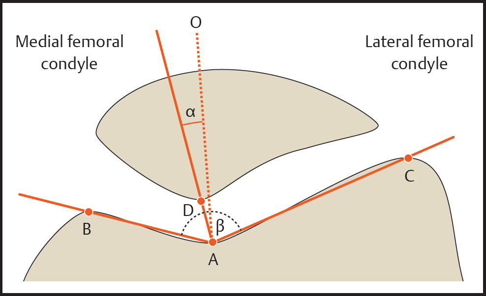


Fig 2


CT scan

Tibial tuberosity-trochlear groove (TT-TG) distance is a measure of lateralization of the tibial tuberosity in relation to the femoral trochlea.

The TT-TG distance represents the radiographic measurement of the quadriceps vector, which represents a lateral force displacement on the patella during knee motion. A line is drawn through the deepest point of the trochlear groove, perpendicular to the posterior femoral condylar tangent (TT). A second line is drawn in parallel to the trochlear line through the most anterior portion of the tibial tubercle (TG). The distance between the 2 lines represented the TT-TG distance. 

It is more than 20 mm in patients with recurrent patellar dislocations, as compared with 13 mm in control subjects (Fig 3).
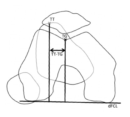
Fig 3.


MRI

It is useful for evaluating loose bodies and assessing the medial patellofemoral ligament (MPFL). Osteochondral lesion and/or bone bruising is commonly seen on the medial patellar facet and the lateral femoral condyle. Tears of the MPFL are usually seen at the medial femoral epicondyle.


Treatment in Adults

Nonoperative

For acute dislocation, a closed reduction is carried out. In majority of patients, spontaneous reduction occurs. The treatment includes the use of NSAIDs, activity modification, and physical therapy.

The indications for non-operative treatment include:

  • First-time dislocation

  • No loose bodies or articular damage

  • No osteochondral fragments

  • Habitual dislocators

  • Patients with connective tissue disease - Ehlers Danlos


Physical therapy focuses on closed chain exercises and quadriceps strengthening exercises. Core hip strengthening and gluteal muscle strengthening helps to improve external rotators of the hip, thus externally rotating the femur and decreasing the Q-angle.

Patellar stabilizing sleeve or "J" brace can be used and patella taping can be carried out. Knee aspiration is carried out if there is tense effusion.


Operative Treatment

The indications for operative treatment include:

  • Osteochondral injury with loose body

  • Chronic instability

  • Failure of nonsurgical treatment


Arthroscopic debridement with removal of loose bodies is done if there are loose bodies or osteochondral damage is seen on imaging. Open reduction internal fixation with screws and pins is carried out if there is sufficient bone available for fixation.

Direct repair of the MPFL can be carried out when there is an acute first-time dislocation with a bony fragment.

MPFL reconstruction with autograft or allograft can be carried out when there is recurrent instability and no malalignment or trochlear dysplasia. Gracillis or semitendinosus is commonly used. The femoral origin can be reliably found (Schottles point). The Schottle point is 1 mm anterior to the posterior cortex line, 2.5 mm distal to the posterior origin of the medial femoral condyle, and proximal to the posterior point of the Blumensaats line. Tensioning of the graft should be done between 60 to 90 degrees of knee flexion. Severe trochlear dysplasia is the most important predictor of residual patellofemoral instability after isolated MPFL reconstruction.

When there is significant malalignment a Fulkerson-type osteotomy is carried out in isolation or in combination with MPFL reconstruction. The osteotomy involves anterior and medial, tibial tubercle transfer with fixation. It is carried out if TT-TG is more than 20mm. It decreases pressure on the lateral patellar facet and trochlea.

Lateral release of the patella has been shown to be ineffective for the treatment of patellar instability. It is used for lateral compression syndrome where there is combined or isolated patellar tilt or excessive tightness after medialization procedure.Usually, the lateral release is combined with a medialization procedure and not done in isolation.

Trochleoplasty to deepen the sulcus of the distal femoral trochlea has limited use due to serious irreversible articular and subchondral injury to the trochlea. It is indicated for abnormal patellar tracking with J sign caused by femoral trochlear dysplasia. The cancellous bone is exposed in the trochlea, and a strip of cortical bone on the edge of the trochlea is elevated. A new trochlea sulcus is created, and the trochlear bone shell is impacted and secured to the new sulcus fixed with staples or sutures.


Pediatric treatment

The principles of treatment in children are the same as in adults, except that the physis must be preserved in children. A tibial tubercle osteotomy should not be done since it will harm the growth plate of proximal tibia. 

Recurrent dislocation rates with nonoperative treatment may be as high as 15-50% at 2-5 years. Recurrence rate is highest in patients who sustain a primary dislocation before the age of 20 years.  

Medial patellar dislocation and medial patellofemoral arthritis can occur as a result of prior patellar stabilization surgery.


Friday 14 October 2022

Adult Spinal Deformity


    Adult Spinal Deformity


                                 Dr. KS Dhillon


Introduction

Adult Spinal Deformity is an idiopathic or degenerative condition of the adult spine. The deformity is in the coronal or sagittal plane. A diagnosis is made with full-length x-rays of the spine. The Initial treatment is usually nonoperative with NSAIDs and physical therapy.  Surgical correction of the deformity is indicated for progressive disabling pain that has not responded to nonoperative management, and in patients with progressive neurological deficits.


Epidemiology

Males and females are equally affected and the mean age is 60 years.

Idiopathic scoliosis is more common in the thoracic spine and degenerative scoliosis occurs more commonly in the lumbar spine.


Etiology

There can be a coronal plane imbalance or sagittal plane imbalance. Sagittal plane imbalance is defined as a radiographic sagittal imbalance of more than 5cm. Coronal plane imbalance is defined as lateral deviation of the normal vertical line of the spine of more than 10 degrees. 

Degenerative scoliosis results from the asymmetric degeneration of disc space and or the facet joints in the spine. When it occurs in the coronal plane it produces scoliosis and in the sagittal plane, it produces kyphosis or lordosis.

Factors that contribute to loss of sagittal plane balance include, preexisting scoliosis, iatrogenic instability, osteoporosis, and degenerative disc disease.


Classification

There are 2 types of coronal deformity. One is idiopathic (residual) adult scoliosis that results from untreated adolescent idiopathic scoliosis and the other is degenerative (de novo) adult scoliosis. Coronal deformity in the adult can be caused by degenerative changes, can be iatrogenic, or can be caused by paralysis.

In idiopathic (residual) scoliosis the curve follows classic curve patterns. It involves more vertebral segments. The curves are large and it is located in the thoracic spine.

Degenerative (de novo) adult scoliosis lacks classic curve patterns. It involves fewer vertebral segments. The curves are smaller and it involves the lumbar spine.


Presentation


Symptoms

Low back pain is the commonest symptom. It is present in 40% to 90% of the patients. The pain is caused by spondylosis, spine instability or the pain can be from the disc. The pain is more severe and recurrent as compared to the general population. 

Spinal stenosis can occur on the concave side of the curve. It can produce neurogenic claudication that would produce pain in the lower extremities and buttocks. Unlike classic claudication, patients with scoliosis and stenosis do not obtain relief of pain with sitting and forward flexion. 

Foraminal and lateral recess stenosis is worse in the concavity of the deformity where there is vertebral body rotation and translation. It can produce radicular leg pain and weakness. 


Physical examination

Examination will show spinal deformity with thoracic prominence when the patient bends forward. There may be associated muscle weakness.


Imaging

Radiographs

Standing scoliosis x-rays in the coronal and sagittal plane with right and left bending films are taken on a full-length long 36-inch cassette. The bending films help assess curve flexibility and the possibility of surgical correction. 

On the AP radiograph, the Cobb angle and coronal balance values are obtained. The coronal balance is calculated using the C7 plumb line (C7PL) and center sacral vertical line (CSVL) (fig 1).

On the lateral radiograph, the sagittal balance is calculated using the C7 plumb line (C7PL). On the lateral radiograph, the pelvic incidence is calculated using sacral slope (SS) and pelvic tilt (PT) values.


Fig 1


CT scan

A CT scan is useful to identify spinal bony abnormalities. It will also help identify bony deformities such as facet arthrosis.


CT myelogram

A CT myelogram is useful for assessing spinal stenosis, bony anatomy, and rotational deformity. 


MRI

In the presence of lower limb pain, an MRI is indicated. It can help identify

central canal stenosis, facet hypertrophy, disc degeneration, pedicular enlargement, and foraminal encroachment. 


Treatment

Nonoperative

In patients with coronal curves of less than 30 degrees, the treatment is conservative because such curves rarely progress. The treatment modalities include NSAIDs, tricyclic antidepressants if there is sleep disturbance, physical therapy, corticosteroid injections, and nerve root blocks to treat pain. Bracing can slow progression and reduce discomfort.


Operative

When surgery is necessary, surgical curve correction with instrumented fusion is carried out. It is usually carried out in patients with curves of more than 50 degrees. Surgery is carried out when there is sagittal imbalance, curve progression, intractable back or radicular pain and when there is

cardiopulmonary decline. Thoracic curves of more than 60 degrees affect pulmonary function tests. Thoracic curves of more than 90 degrees can predispose to mortality. Sometimes surgery is carried out for cosmesis.

Posterior only curve correction and instrumented fusion is carried out in patients with thoracic curves of more than 50 degrees and most double structural curves of more than 50 degrees. 

A combined anterior and posterior curve correction with instrumented fusion is carried out in patients with isolated thoracolumbar and isolated lumbar curves. Extremely rigid curves will require anterior release.

The aim of surgery is to restore spinal balance, relieve pain and obtain a solid fusion. Worse outcomes is seen in patients with baseline depression and obesity.

The proximal fusion level extends to a neutral and horizontal vertebra above the main curve. The distal fusion extends to L5 if there is no pathology at L5/S1. There is a high failure rate if instrumentation does not extend to the sacrum if there is pathology at L5/S1.

The fusion should be extended to the sacrum when there is pathology at 

L5-S1 such as spondylolisthesis, spondylolysis, and facet arthrosis.

A prior laminectomy at L5-S1 would also be an indication for extending the fusion to the sacrum. 

A concomitant anterior release and anterior column support through an anterior approach may be needed for better correction of the deformity. The advantage of the long fusion construct is increased stability of the fusion construct and it is less likely to fail.  

The disadvantages include an increased risk of pseudoarthrosis, increased surgical time, increased reoperation rate, increased risk of sacral insufficiency fractures, and altered postoperative gait. 

The fusion can be extended to the ilium (sacropelvic fusion) if the sacrum is included in fusion involving more than 3 levels.

In osteoporotic patients, cement augmentation can be used. The cement is injected through a fenestrated tap at the end vertebra followed by pedicle screw insertion. 

In patients with severe angular deformities, osteotomies are useful to regain sagittal balance. Thirty degrees or more correction can be obtained through Smith-Petersen or pedicle subtraction osteotomies. When osteotomy is carried out intraoperative neuromonitoring is required.

A Smith-Petersen osteotomy (SPO) is carried out for mild to moderate sagittal imbalance that requires correction of up to 10 degrees per level of osteotomy. The prerequisites are that there should be no anterior fusion at the level of osteotomy and there should be adequate disc height and mobility. The correction is at the level of the disc. There is more correction at the lumbar spine due to greater disc height and mobility. The correction is less in the thoracic spine due to lesser disc height and mobility.

In patients with severe sagittal imbalance of more than 12cm requiring correction of 30 degrees to 35 degrees in the lumbar spine, and 25 degrees in the thoracic spine, where anterior fusion is present, pedicle subtraction osteotomy (PSO) is performed. The correction is at the level of the vertebral body and not at the disc.

Vertebral column resection becomes necessary when there is severe sagittal imbalance requiring correction of up to 45 degrees. It is also necessary when there is rigid angular thoracic spine kyphosis, such as that associated with tumors, fractures, or infection. Severe rigid scoliosis and congenital kyphosis would require hemivertebrae resection in the thoracic or lumbar spine.

Anterior procedures are carried out for rigid large curves of more than 70 degrees. Rigid curves are those with no flexibility on side-bending films.

Anterior procedures are also carried out for isolated lumbar or thoracolumbar curves and for anterior interbody fusion at L5/S1 when fusing to the sacrum. Anterior release and fusion are usually combined with posterior instrumentation and fusion.

The disadvantages of anterior procedures are that the surgery is longer if performed on the same day and the complication rates are higher and they are medically more stressful. The advantages are that they increase the stability of L5-S1 long fusion constructs and helps to restore and maintain sagittal and coronal balance. 


Surgical Complications

The overall complication rate of adult spinal deformity surgery is about 13.5%. There are 10% major complications that often irreversibly affect the long-term health of the patient. The complication rate is significantly higher when osteotomies, revision procedures, and combined anterior/posterior surgery is performed.


The complications include:  


1. Pseudoarthrosis    

The incidence of pseudoarthrosis following the surgery is between 5% and 25%. The most common surgical technique associated with pseudoarthrosis is posterior only fusion. The incidence is about 15%. Pseudoarthrosis is most commonly seen at the L5-S1 junction and at the thoracolumbar junction. 

The risk factors for pseudoarthrosis include age more than 55 years, hip arthritis, smoking, kyphosis more than 20 degrees, positive sagittal balance of more than 5cm, thoracoabdominal approach, and incomplete lumbopelvic fixation. 


2. Dural tear

The incidence of dural tears is about 2.9%.


3. Infection

The incidence of deep wound infection is about 1.5% and superficial wound infection about 0.9%. The risk of infection increases with smoking, diabetes, increasing age, and revision surgery.     


4. Implant complication 

Implant failure is more likely to occur in bone with lowest ratio of cortical to cancellous bone. Implant failure is the most common cause of reoperation  (38.2%) followed by infection (11.8%).   


5. Neurologic deficits

Acute neurological deficits can occur in about 1.0% of the patients intraoperatively during deformity correction maneuver. If it is identified on 

neurophysiologic monitoring, the instrumentation should be removed, and a wake-up test done. Delayed neurological deficits occurs in about 0.5% of the patients.

Acute neurological deficits can occur due to nerve root injury caused by screw malposition and corrective maneuver.


6. Epidural hematoma 

Epidural haematoma occurs in about 0.2% of the patients. 


7. Pulmonary embolus 

Pulmonary embolism can be seen in about 0.2% of the patients.


8. Deep venous thrombosis 

Deep venous thrombosis is seen in about 0.2% of the patients.


9. Deaths 

Death as a complication is seen in about 0.3% of the patients.


Prognosis

Worse prognosis is seen in patients in whom the symptoms progress to the side of curve convexity and in patients with sagittal plane imbalance. A sagittal plane balance is the most reliable predictor of clinical symptoms in adult patients with spinal deformity.           

Thoracic curves progress more than lumbar curves, lumbar curves more than thoracolumbar curves and thoracolumbar curves more than double major curves. Right thoracic curves progress about 1 degree per year, right lumbar curves about 0.5 degrees per year and thoracolumbar curves about 0.25 degrees per year.

Curves that are less than 30 degrees rarely progress. Curves that are more than 50 deg commonly progress. In patients with preexisting rotational changes progression also occurs.


Tuesday 4 October 2022

Flexor Tendon Injuries Of The Hand


      Flexor Tendon Injuries Of The Hand


                           Dr. KS Dhillon


Anatomy of Flexor tendons


Muscles

The flexor digitorum profundus arises from the proximal 3/4 of medial and anterior surfaces of the ulna and interosseous membrane. It is inserted at the base of the distal phalanx of the fingers. The medial part (slips to little and ring finger) is innervated by the ulnar nerve (C8, T1) and the lateral part (slips to index and middle finger) is innervated by the anterior interosseous nerve (C8, T1). The ulnar and anterior interosseous arteries provide the blood supply. It functions as a flexor of the distal interphalangeal (DIP) joint and it assists with proximal interphalangeal (PIP) and metacarpophalangeal (MCP) flexion.

Flexor digitorum superficialis (FDS) originates from the medial epicondyle of the humerus, ulnar collateral ligament, coronoid process of the ulna, and superior half of the anterior border of the radius. It is inserted on the bodies of the middle phalanges of fingers. It functions as a flexor of the middle phalanges at the PIP joints of the fingers. It also flexes the proximal phalanges at MCP joint. It is innervated by the median nerve (C7, C8, T1). The arterial supply is provided by the ulnar artery. The FDS to the small finger is absent in 25% of people. 

The flexor pollicis longus (FPL) originates from the anterior surface of the radius and adjacent interosseous membrane. It is inserted on the base of the distal phalanx of the thumb. It flexes the phalanges of the thumb. It is located within the carpal tunnel as the most radial structure. It is innervated by the anterior interosseous nerve (C8, T1). The blood supply is provided by the anterior interosseous artery.

The flexor pollicis brevis has 2 heads. The superficial head originates from the flexor retinaculum and tubercles of scaphoid and trapezium. The deep head orginates from the trapezoid and capitate (more medial than superficial head). The flexor pollicis brevis is inserted on the lateral side of base of proximal phalanx of thumb. It functions as a thumb flexor. The superficial head is innervated by the recurrent branch of the median nerve (C8, T1) and deep head is innervated by the deep branch of ulnar nerve.

The arterial supply is from the superficial palmar branch of the radial artery. 

The flexor carpi radialis (FCR) originates from the medial epicondyle of the humerus and is inserted on the base of the 2nd metacarpal. It lies close to the median nerve. It flexes and abducts the hand at the wrist. It is innervated by the median nerve (C6, C7). The blood supply is provided by the ulnar artery. 

The flexor carpi ulnaris (FCU) originates from the medial epicondyle of the humerus, the olecranon, and the posterior border of the ulna. It is inserted on the pisiform bone, hook of hamate bone, and the 5th metacarpal bone. It flexes and adducts the hand at the wrist. It is innervation by the ulnar nerve (C7, C8). The arterial supply is from the ulnar artery.


Camper chiasm

The tendinous chiasm is situated where tendons of flexor digitorum superficialis and flexor digitorum profundus muscles cross each other. 

The FDS tendon enters the A1 pulley and divides into 2 equal halves that rotate laterally and then dorsally. The 2 slips rejoin deep to the FDP tendon over the distal aspect of the proximal phalanx and the palmar plate of the PIP joint at the camper’s chiasm. The 2 slips then insert separately on the volar aspect of the middle phalanx (fig 1).


Fig 1. 


Pulley system  

There are 5 annular pulleys (A1 to A5) in each finger. They are thicker and stiffer than the cruciate pulleys. The A2 and A4 pulleys arise from the periosteum and are important pulleys to prevent flexor tendon bowstringing. The A1, A3, and A5 arise from the volar plate and overlie the MP, PIP, and DIP joints respectively (fig 2). The A1 pulley is most commonly involved in a trigger finger.

Fig 2.


There are 3 cruciate pulleys (C1 to C3). They are situated at the level of the joints (fig 2). They function to prevent sheath collapse and expansion during digital motion. They facilitate approximation of annular pulleys during flexion. 

The thumb contains 3 annular pulleys (A1, Av, A2). The A2 contributes least to the arc of motion of the thumb (fig 3). The Av oblique pulley is the most important pulley to prevent flexor tendon bowstringing (along with the A1 pulley). The oblique pulley (3-5mm) originates at the proximal half of the proximal phalanx. It facilitates full excursion of flexor pollicis longus (FPL)  and prevents bowstringing of FPL. Bowstringing will occur if both A1 and oblique pulleys are cut. The blood supply comes from 2 sources. One is by 

diffusion through synovial sheaths and direct vascular perfusion outside of synovial sheaths. 


Fig 3


Zones of flexor tendon injuries (fig 4)

Zone 1 is distal to the FDS insertion. Injury to the FDP in this zone produces a Jersey finger.  A Jersey finger is a traumatic flexor tendon injury caused by an avulsion injury of the FDP from the insertion at the base of the distal phalanx. On clinical examination, the finger lies in slight extension at the DIP relative to other fingers in the resting position. X-rays may show a bony avulsion if present. Treatment is direct tendon repair or open reduction and internal fixation depending on the presence and size of a bony avulsion.

Zone II extends from the FDS insertion to the distal palmar crease/proximal A1 pulley. In this zone, FDP and FDS are in the same tendon sheath (both can be injured within the flexor retinaculum). The tendons can retract if the vincula are disrupted. The treatment is by direct tendon repair followed by early range of movement exercises (Duran, Kleinert). Passive finger flexion and active extension to an orthosis is carried out. Historically this zone had very poor results but results have improved due to advances in postoperative motion protocols.

Zone III is situated in the palm. It extends from the A1 pulley to the distal aspect of the carpal ligament. Injuries in this zone are often associated with neurovascular injury which carries a worse prognosis. The treatment is by direct tendon repair. Good outcome from direct repair can be expected due to the absence of retinacular structures provided there is no neurovascular injury. An A1 pulley release may be needed to avoid impingement of the repaired tendon on the pulley.

Zone IV is situated in the carpal tunnel. The tendon surgery is often complicated by postoperative adhesions due to close quarters and synovial sheath of the carpal tunnel. The treatment is by direct tendon repair. The 

transverse carpal ligament should be repaired in a lengthened fashion if tendon bowstringing is present.

Zone V extends from the carpel tunnel to the forearm. The tendon injury is 

often associated with neurovascular injury which carries a worse prognosis.

The treatment is by direct tendon repair.

In the thumb, there are three zones (TI,TII,TIII). The outcome of thumb tendon injuries is different from finger injuries. The early motion protocols do not improve the long-term results. There is a higher re-rupture rate as compared to flexor tendon repair in fingers. The treatment is by direct end-to-end repair of FPL. Zone III should be avoided to prevent injury to the recurrent motor branch of the median nerve. 

Fig 4


Clinical Presentation

Symptoms of flexor tendon injury include a loss of active flexion strength and motion of the involved digit. Normally passive wrist flexion and extension allows for assessment of the tenodesis effect. Normally wrist extension causes passive flexion of the digits at the MCP, PIP, and DIP joints. Maintenance of extension at the PIP or DIP joints with wrist extension indicates flexor tendon discontinuity. Active PIP and DIP flexion is tested in isolation for each digit. 

A neurovascular examination is important given the close proximity of flexor tendons to the digital neurovascular bundles.

X-rays are taken to exclude associated fractures.


Treatment


Nonoperative

Partial lacerations of less than 60% of tendon width can be treated non operatively with wound care and early range of motion exercises.


Operative

Lacerations of more than 60% of tendon width are treated surgically with flexor tendon repair and controlled mobilization.

In patients with failed primary repair and in patients with chronic untreated injuries flexor tendon reconstruction and intensive postoperative rehabilitation are necessary.  

In patients with chronic FPL rupture, a FDS of the ringer finger is transferred to the thumb in a single-stage procedure.


Techniques of tendon repair

The indications for flexor tendon repair include a more than 75% laceration of the tendon and equal to or more than 50-60% laceration with triggering. An epitendinous suture at the laceration site is sufficient. There are no     

benefits of adding a core suture.

Fundamentals of repair include easy placement of sutures in the tendon,

secure suture knots, smooth juncture of the tendon ends, minimal gapping at the repair site, minimal interference with tendon vascularity, and sufficient strength throughout healing to permit application of early motion stress to the tendon.

The repair should be performed within three weeks of injury, ideally within 2 weeks. Delayed treatment leads to difficulty due to tendon retraction.

Incisions should always cross flexion creases transversely or obliquely to avoid contractures. The tendon handling should be atraumatic to minimize adhesions.

The number of suture strands that cross the repair site is more important than the number of grasping loops. There is a linear relationship between the strength of the repair and the number of sutures crossing the repair. Four to six strands provide adequate strength for early active motion. A high-caliber suture material increases strength and stiffness and decreases gap formation. Locking loops also decrease gap formation. The site of ideal suture placement is 10mm from the cut edge.    

Circumferential epitendinous suture improves tendon gliding by reducing the cross-sectional area. It improves the strength of repair by adding 20% to tensile strength. It allows for less gap formation. Simple running suture is recommended. 

A repair of the sheath theoretically improves tendon nutrition through the synovial pathway. Clinical studies, however, show no difference with or without sheath repair.

Tendon repairs are weakest between postoperative day 6 and day 12. The 

repair usually fails at suture knots. Repair site gaps of more than 3mm are associated with an increased risk of repair failure. Adhesion formation is more common with zone 2 injuries.


Flexor tendon reconstruction    

For flexor tendon reconstruction the skin must be supple, sensation intact and there should be adequate vascularity. The passive range of movements of the adjacent joints must be full. The procedure can be a single-stage or two-stage procedure.

A single-stage procedure is only performed if the flexor sheath is pristine and the digit has full ROM. 

There are 2 types of two-stage procedures:

1. Hunter-Salisbury

Here in stage I a tendon spacer is placed to create a favorable tendon bed. In stage II, about 3-4 months later, the tendon spacer is retrieved and a tendon graft is placed through the mesothelium-lined pseudosheath. 

Proximally pulvertaft weave is done and distally end-to-end tenorrhaphy is carried out. Postoperatively a Kleinert´s splint is applied and after 24 hours finger exercises are started.

2. Paneva-Holevich  

In stage I tendon spacer is placed in the flexor sheath, pulleys are reconstructed (as needed), and a loop between the proximal stumps of FDS and FDP is created in the palm. 

In stage II the tendon spacer is retrieved, FDS is cut proximally and reflected distally through the pseudosheath and either attached directly to the FDP stump or secured with a button.

The most commonly used tendon for grafting is the palmaris longus. It is however absent in 15% of the population. If a longer graft is necessary the plantaris is used. It is absent in 19% of the population. Other grafts available include extensor digitorum longus to 2nd-4th toes, extensor indicis proprius, flexor digitorum longus to 2nd toe, and FDS.

One pulley should be reconstructed proximal and distal to each joint. Pulley reconstruction should be done first if a tendon graft is being used. 

The A2 pulley is important. It is unclear if A4 reconstruction is absolutely necessary. For graft material, the extensor retinaculum synovialized pulley surface provides the least gliding resistance. The palmaris, plantaris, FDS, and flexor tendon allograft can be used as graft material. 

First, all scar dorsal to the flexor tendon is excised.

In the encircling technique or around-the-bone technique, a single-loop (Bunnell) or triple loop (Okutsu) technique can be used. The triple loop technique is biomechanically strongest construct.

There are several techniques for the nonencircling reconstruction. These include ever-present-rim (Kleinert), belt-loop (Karev), extensor retinaculum (Lister), and the palmaris longus transplantation through volar plate (Doyle and Blythe) techniques.


Tenolysis

Tenolysis is indicated when there is localized tendon adhesions with minimal to no joint contracture and full passive finger movements. It may also be required if there is a discrepancy between active and passive motion of the fingers after therapy. Tenolysis is carried out after 3 months when the soft tissues have stabilized and there is full passive motion of all joints. While doing tenolysis one has to be careful to preserve A2 and A4 pulleys. Intensive physiotherapy is carried out post-operatively.


Postoperative rehabilitation after tendon repair

Controlled mobilization after the surgery has been the major reason for improved results with tendon repair, especially in zone II. It leads to improved tendon healing biology. It also limits restrictive adhesions and leads to increased tendon excursion. 

Immobilization using a splint with wrist and MCP joints in flexion and IP joints in extension is necessary for children and non-compliant patients.  

Early passive motion is started using the Duran protocol (active finger extension with patient-assisted passive finger flexion and static splint) or the Kleinert protocol (active finger extension with dynamic splint-assisted passive finger flexion).

Early active motion is also started with moderate force with a dorsal blocking splint limiting wrist extension. 


Complications

There are several complications that arise following tendon repairs.

1. Tendon adhesions

Tendon adhesions is the most common complication following flexor tendon repair. The risk is highest with zone 2 injuries. The adhesions are initially treated with physical therapy. If physical therapy does not work tenolysis is performed. Tenolysis is performed 4-6 months after tendon repair if there is a significant loss of excursion.

2. Rerupture  

The rerupture rates are between 15-25%. If the scar is less than 1cm, the scar is resected and primary repair is carried out. If the scar is more than 

1cm the scar is excised and a tendon graft is performed. If the sheath is collapsed, a Hunter rod is placed, and staged grafting is performed.

3. Joint contracture

A joint contracture is seen in about 17% of the patients. A contracture release has to be carried out sometimes.

4. Swan-neck deformity 

Clinical examination reveals resting hyperextension of the PIP joint combined with resting flexion of the DIP joint of the involved digit. It is initially treated with a course of PIP splinting to prevent hyperextension. In progressive cases, volar plate advancement with central slip tenotomy is performed. 

5. Trigger finger  

Trigger finger is the inhibition of smooth tendon gliding due to mechanical impingement at the level of the A1 pulley. The triggering causes pain, clicking, catching, and locking of the finger. Physical examination shows the presence of active triggering and tenderness at the A1 pulley. Treatment consists of anti-inflammatory medications and steroid injections. In resistant cases surgical release may be necessary.

6. Lumbrical plus finger

Lumbrical plus finger is characterized by extension of the IP joints while attempting to flex the fingers. Clinical examination will show extension of the IP joints of one digit when the fingers are flexed to make a fist. When the symptoms are minimal no treatment is required. When the symptoms affects patients activities surgical treatment is carried out with tenodesis of FDP to terminal tendon or with a lumbrical release.   

7. Quadrigia

It is most commonly caused by a functional shortening of the FDP tendon due to over-advancement of the FDP during tendon repair. A more than 1 cm advancement is associated with quadriga. It is characterized by an active flexion lag in fingers adjacent to a digit with a previously repaired FDP tendon. Clinical examination will show the inability to fully flex the fingers of the hand adjacent to the injured finger. If there are minimal symptoms no treatment is reuired. If it affects the patients activities than surgery is carried to release the FDP tendon.