Achilles Tendon Rupture
Dr. KS Dhillon
Introduction
The Achilles tendon is the strongest tendon in the human body [1]. It is the most common tendon rupture in the lower extremities. The injury commonly occurs in adults in their third to fifth decade of life [2]. Acute ruptures usually present with sudden onset of pain associated with a snapping or audible pop heard at the site of injury. Patients often describe the sensation of being kicked in the back of the lower leg. The injury causes significant pain and disability.
These injuries typically occur in individuals who are only active intermittently (the "weekend warrior" athletes). The injury is misdiagnosed as an ankle sprain in 20% to 25% of patients. Ten percent of the patients give a history of prodromal symptoms. The known risk factors for tendon rupture include prior intratendinous degeneration, fluoroquinolone use, steroid injections, and inflammatory arthritides [3,4,5,6].
Anatomy
The Achilles tendon is the strongest and largest tendon in the body. The tendinous fibers of the gastrocnemius originating from the distal femur and those of the soleus muscle originating from the proximal tibia coalesce to form the Achilles tendon and the tendon inserts on the posterior calcaneal tuberosity. The tendon is approximately 15cm in length. It travels distally and twists approximately 90° internally so that its initial anterior fibers of the gastrocnemius insert laterally and the initial posterior fibers of the soleus insert on the medial aspect of the Achilles tendon. The Kager's fat pad that is located anterior to the Achilles tendon protects blood vessels entering the tendon.
The tendon has no tendon sheath but it has a highly vascularized paratenon that acts as a conduit for the vasculature of the tendon and it facilitates tendon gliding between the subcutaneous tissue and posterior fascia. The proximal and distal sections of the tendon are supplied by the posterior tibial artery and the midsection which is 2 to 6 cm from the insertion point is supplied by the peroneal artery. Since the midsection receives a relatively poor blood supply, it is most vulnerable to degeneration and rupture.
Etiology
There are several causes for the rupture of the tendon Achilles and these include sudden forced plantar flexion of the foot, direct trauma to the tendon, and long-standing tendinopathy or intratendinous degeneration. Sports that are commonly associated with Achilles tendon rupture include tennis, diving, basketball, and track events. Poor conditioning before exercise, prolonged use of corticosteroids, overexertion, and the use of quinolone antibiotics increases the risk of tendon rupture. The rupture usually occurs about two to four cm proximal to the calcaneal insertion of the tendon. In those who are right-handed, the left Achilles tendon is most likely to rupture and vice versa [7,8,9].
The cause of Achilles tendon injury is multifactorial. The injury is most often seen in gymnasts, cyclists, runners, and volleyball players. In cyclists, the combination of low saddle height and extreme dorsiflexion of the ankle during pedaling may be a factor responsible for the injury.
There are systemic diseases that can be associated with Achilles tendon injuries and these include [10]:
- Chronic renal failure
- Collagen deficiency
- Diabetes mellitus
- Gout
- Infections
- Lupus
- Parathyroid disorders
- Rheumatoid arthritis
- Thyroid disorders
There are foot problems that can increase the risk of Achilles tendon injuries and these include [10]:
- Cavus foot
- Insufficient gastrocsoleus flexibility and strength
- limited ability to perform ankle dorsiflexion
- Tibia vara
- Varus alignment with functional hyperpronation
Achilles tendon rupture is often more common in people with O blood group. Furthermore, anyone with a family history is also at a higher risk of developing Achilles tendon rupture.
Epidemiology
The incidence of Achilles tendon rupture varies in the literature. Recent studies report a rate of 18 patients per 100,000 patient population annually [10]. In athletes, the incidence of achilles tendon injuries ranges from 6% to 18%. Football players are the least likely to develop this problem compared to tennis players and gymnasts. About a million athletes suffer from Achilles tendon injuries each year [11].
The true incidence of Achilles tendinosis is not known. The reported incidence rates are 9% in dancers, 7% to 18% in runners, 5% in gymnasts, 2% in tennis players, and less than 1% in American football players.
The incidence of Achilles tendon injuries is on the increase because more people are participating in sports.
Achilles tendon injuries are more common in males (6:1, male-female ratio), and this is probably related to greater participation in sporting activities. Most injuries occur between the third and fifth decade of life.
Pathophysiology
Achilles tendonitis is usually not associated with primary prostaglandin mediated inflammation. There is neurogenic inflammation with the presence of calcitonin gene-related peptide and substance P. Histopathological examination shows thickening and fibrin adhesions of the tendon with the occasional disarray of the fibers [10].
Neurovascularization is usually seen in the degenerating tendon. Tendon rupture is usually the terminal event of this degenerative process. After rupture, type 111 collagen is the major collagen manufactured, which suggests that the repair process is incomplete. Studies in animals show that if there is more than 8% stretching of the original length, tendon rupture is likely.
The proximal part of the tendon receives its blood from the muscle bellies connected to the tendon. Blood supply to the distal segment of the tendon comes from the tendon-bone interface.
History and Physical Examination
Patients with tendon Achilles rupture usually present with acute, sharp pain in the region of the Achilles tendon. Physical examination shows that the patient is unable to stand on the toes. The ankle plantar flexion is very weak. Palpation may show a discontinuity of the tendon. There are signs of bruising around the posterior ankle.
The following clinical test can help in the diagnosis of tendon Achilles rupture:
1. Thompson test: Also known as the “calf squeeze test,” it is an accurate test to detect Achilles tendon rupture. The patient is made to lie prone with the knee flexed to 90 degrees. The gastrocnemius is then squeezed and if the ankle does not plantarflex then the test is positive and it indicates the presence of a rupture.
2. Matles test (Fig 1): The patient is placed in the prone position with the knees flexed at 90 degrees. In a positive Matles test the affected foot is in an increased dorsiflexed position instead of a resting plantar flexed position when compared to the contralateral limb.
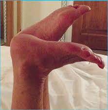
Fig 1- Matles test
Evaluation
An x-ray of the ankle can be done to rule out a fracture of the posterior calcaneus and exclude other pathology. An ultrasound can be useful to determine partial from complete tears. An MRI will be useful when the clinical findings are equivocal and in patients with chronic ruptures. The MRI will show acute rupture with retracted tendon edges.
Management of the rupture
The initial treatment of Achilles tendon rupture is rest, elevation, pain control, and functional bracing. The debate surrounding the potential benefits versus risks of surgical intervention still continues. Several studies have demonstrated good functional results and patient satisfaction with both operative and nonoperative treatment modalities.
Healing rates with serial casting/functional bracing are the same as compared to surgical repair of the tendon. However, return to work may be slightly prolonged in patients treated nonoperatively. All patients require both physical and orthotic therapy to help strengthen the muscles and improve the range of motion of the ankle [5,12,13].
Rehabilitation is critical to regaining optimal ankle function. While there remains debate regarding the optimal treatment modality, the general consensus includes the following:
1. Patients with relatively sedentary lifestyle and those with significant medical comorbidities are usually recommended to have nonoperative treatment.
2. Those with soft tissue/skin integrity problems are also advised nonoperative treatment.
3. The patient and surgeon discussion should include a detailed discussion about the current literature reporting satisfactory outcomes with both treatment plans.
3. Possibility of quicker return to work with operative intervention should be made known to the patient.
4. The plantar flexion strength is the same on long term follow up with both treatments.
5. There is an increased risk of re-rupture and/or re-injury with nonoperative treatment as compared to operative treatment.
6. The complication rates are higher with operative treatment.
There are several techniques for repair of the Achilles tendon, but all involve approximation of the torn ends. Sometimes the repair is reinforced by the gastrocsoleus aponeurosis or the plantaris tendon. Overall, the healing rates between casting and surgical repair are quite similar.
The claims about an early return to activity after surgery are now being questioned. If a cast is applied, it should remain for at least 6-12 weeks. There are several benefits of a non-surgical approach and that include no hospital admission costs, no wound complications and no risk of anesthesia.
New studies show equivalent rates of re-rupture after functional rehabilitation and operative repair [14].
Operative treatment can include:
1. Open end-to-end achilles tendon repair. It is indicated in patients with acute ruptures (approximately <6 weeks).
2. Percutaneous Achilles tendon repair. It is indicated where there are concerns over cosmesis of traditional scar. With this technique there is a higher risk of sural nerve injury. There is a lesser risk of infection and wound complications as compared with open repair.
3. Reconstruction with VY advancement. This is indicated in patients with chronic ruptures with defects of less than 3cm.
4. Flexor hallucis longus transfer with or without VY advancement of gastrocnemius. It is indicated in patients with chronic ruptures with a more than 3cm defect. It requires a functioning tibial nerve.
Complications
Complications of operative treatment of acute Achilles tendon rupture include infection, sural nerve injury, rerupture, deep vein thrombosis, and hypertrophic scars. Hence, operative treatment may not be appropriate for patients with diabetic mellitus, and peripheral vascular disease and in low-demand patients.
Infection
The most serious complication following open tendon repair is infection. Infection and wound problems occur after surgery and the incidence of these complications is about 12.5% [15,16].
To prevent an infection, superficial dissection should be avoided during incision and the synovial tissue envelope should be restored as much as possible before the paratenon is repaired. Minimal number of sutures must be used to obviate delayed infection around the subcutaneous suture knot. Absorbable sutures are preferable to nonabsorbable sutures, which increase the risk of delayed infection or irritation.
Calf Muscle Weakness
Even without proper healing of a ruptured Achilles tendon, individuals are able to walk. A permanent functional deficit, however, remains. The ultimate goal of treatment of patients with tendoachilles rupture is to prevent residual calf muscle weakness. The ability to perform a single heel raise is a valid indicator of calf muscle strength. Most patients with a neglected tear are unable to perform a single heel raise [17,18].
Rerupture
Rerupture of the tendon Achilles has been reported after surgical repair. Rettig et al [19] reported a postoperative rerupture rate of 4.5% in their patients, and 16.6% of this occurred in those aged 30 years or younger. Care must be taken during aggressive rehabilitation in younger patients. Reito et al [20] reported a rerupture rate of 7.1% in 210 patients with acute Achilles tendon rupture after conservative treatment. This complication occurred within 12 weeks after treatment in most cases. They suggested extra care should be taken in the first month after nonoperative treatment. Young et al [21] found that 9 of the total 12 reruptures (75%) occurred within 3 months after surgery and that there was no association between the rerupture rate and the repair method.
Prognosis
Generally, patients will resume normal ambulation within 12.5 to 18 weeks after an acute rupture of the Achilles tendon [22]. Without doubt early weight bearing and rehabilitation contributes to improved prognosis [14,22,23].
Patients are usually advised against running and non-contact sports for 16 to 20 weeks following the injury [24]. Van Sterkenburg et al [25] suggested the following criteria for return to running:
- The ability to perform repetitive single heel raises and toe walking
- Equal to or less than 25% calf strength deficit compared to the normal side at 12 weeks after injury.
According to Olsson et al [26], the heel raise ability is an important indicator of general level of tendon healing. Their study showed that 40 out of 81 patients (49%) with acute Achilles tendon ruptures were unable to perform a single heel raise at 12 weeks after the injury.
In a study by Ryu et al [27] 87 of their 112 patients with acute Achilles tendon ruptures had difficulty with a single heel raise at 3 months after open tenorrhaphy followed by early rehabilitation. However, at 6 months postoperation all patients were able to raise the heel.
McCormack and Bovard [28] carried out a systematic review and meta-analysis of randomised controlled trials and found that patients had a 10% to 30% calf strength deficit on the injured side compared to the uninjured side in patients with acute Achilles tendon tears.
Ryu et al [27] also found that in patients who were able to perform single heel raises and sports after operative repair of acute tears and early rehabilitation, the calf circumference decreased by an average of 1.6 cm on the injured side. The isokinetic flexion peak torque deficit at 30°/sec was 16% (range, 0% to 21%) on the injured side as compared with the uninjured side.
Conclusions
Acute Achilles tendon ruptures must be differentiated from ruptures that occur due to chronic degeneration of the tendon. Acute rupture of a healthy tendon can be successfully treated either conservatively or surgically. Rehabilitation is a crucial component of treatment following both methods of treatment. The patient's adherence to rehabilitation should be taken into consideration when treating patients with ruptured tendon Achilles. Rehabilitation during the first 6 months after injury is very important. The focus of rehabilitation is to prevent rerupture during the first 2 months and improving calf muscle strength between 2 months and 3 months after injury. Between 3 months and 6 months after injury, rehabilitation efforts are directed toward return to sports through strengthening and proprioceptive exercises. During rehabilitation care should be taken not to cause hyperdorsiflexion of the ankle to prevent calf muscle weakness.
Surgical treatment can be associated with complications. Heel raise ability is an important indicator of general level of tendon healing. By 6 months post operation all patients are usually able to do a heel raise.
In patients with Achilles tendon tear, there is a 10% to 30% calf strength deficit on the injured side compared to the uninjured side. The calf circumference is also decreased by an average of 1.6 cm on the injured side.
References
- Järvinen TA, Kannus P, Paavola M, Järvinen TL, Józsa L, Järvinen M. Achilles tendon injuries. Curr Opin Rheumatol. 2001 Mar;13(2):150-5. doi: 10.1097/00002281-200103000-00009. PMID: 11224740.
- Järvinen TA, Kannus P, Maffulli N, Khan KM. Achilles tendon disorders: etiology and epidemiology. Foot Ankle Clin. 2005 Jun;10(2):255-66.
- Carmont MR. Achilles tendon rupture: the evaluation and outcome of percutaneous and minimally invasive repair. Br J Sports Med. 2018 Oct;52(19):1281-1282.
- Noback PC, Freibott CE, Tantigate D, Jang E, Greisberg JK, Wong T, Vosseller JT. Prevalence of Asymptomatic Achilles Tendinosis. Foot Ankle Int. 2018 Oct;39(10):1205-1209.
- Haapasalo H, Peltoniemi U, Laine HJ, Kannus P, Mattila VM. Treatment of acute Achilles tendon rupture with a standardised protocol. Arch Orthop Trauma Surg. 2018 Aug;138(8):1089-1096.
- Yasui Y, Tonogai I, Rosenbaum AJ, Shimozono Y, Kawano H, Kennedy JG. The Risk of Achilles Tendon Rupture in the Patients with Achilles Tendinopathy: Healthcare Database Analysis in the United States. Biomed Res Int. 2017;2017:7021862.
- Alušík Š, Paluch Z. [Drug induced tendon injury]. Vnitr Lek. 2018 Winter;63(12):967-971.
- Ahmad J, Jones K. The Effect of Obesity on Surgical Treatment of Achilles Tendon Ruptures. J Am Acad Orthop Surg. 2017 Nov;25(11):773-779.
- Egger AC, Berkowitz MJ. Achilles tendon injuries. Curr Rev Musculoskelet Med. 2017 Mar;10(1):72-80.
- Shamrock and Varacallo. Achilles Tendon Rupture at https://www.ncbi.nlm.nih.gov/books/NBK430844/.
- Maffulli N, Via AG, Oliva F. Chronic Achilles Tendon Rupture. Open Orthop J. 2017;11:660-669.
- Kanchanatawan W, Densiri-Aksorn W, Maneesrisajja T, Suppauksorn S, Arirachakaran A, Rungchamrussopa P, Boonma P. Hybrid Achilles Tendon Repair. Arthrosc Tech. 2018 Jun;7(6): e639-e644.
- Westin O, Svensson M, Nilsson Helander K, Samuelsson K, Grävare Silbernagel K, Olsson N, Karlsson J, Hansson Olofsson E. Cost-effectiveness analysis of surgical versus non-surgical management of acute Achilles tendon ruptures. Knee Surg Sports Traumatol Arthrosc. 2018 Oct;26(10):3074-3082.
- Willits K, Amendola A, Bryant D, Mohtadi NG, Giffin JR, Fowler P, Kean CO, Kirkley A. Operative versus nonoperative treatment of acute Achilles tendon ruptures: a multicenter randomized trial using accelerated functional rehabilitation. J Bone Joint Surg Am. 2010 Dec 1;92(17):2767-75. doi: 10.2106/JBJS.I.01401. Epub 2010 Oct 29. PMID: 21037028.
- Soroceanu A, Sidhwa F, Aarabi S, Kaufman A, Glazebrook M. Surgical versus nonsurgical treatment of acute Achilles tendon rupture: a meta-analysis of randomized trials. J Bone Joint Surg Am. 2012;94(23):2136–2143.
- Kadakia AR, Dekker RG, 2nd, Ho BS. Acute Achilles tendon ruptures: an update on treatment. J Am Acad Orthop Surg. 2017;25(1):23–31.
- Elias I, Besser M, Nazarian LN, Raikin SM. Reconstruction for missed or neglected Achilles tendon rupture with V-Y lengthening and flexor hallucis longus tendon transfer through one incision. Foot Ankle Int. 2007;28(12):1238–1248.
- Yasuda T, Shima H, Mori K, Kizawa M, Neo M. Direct repair of chronic Achilles tendon ruptures using scar tissue located between the tendon stumps. J Bone Joint Surg Am. 2016;98(14):1168–1175.
- Rettig AC, Liotta FJ, Klootwyk TE, Porter DA, Mieling P. Potential risk of rerupture in primary achilles tendon repair in athletes younger than 30 years of age. Am J Sports Med. 2005;33(1):119–123.
- Reito A, Logren HL, Ahonen K, Nurmi H, Paloneva J. Risk factors for failed nonoperative treatment and rerupture in acute Achilles tendon rupture. Foot Ankle Int. 2018;39(6):694–703.
- Young KW, Lee HS, Park SC. Rerupture risk after tenorrhaphy for Achilles tendon rupture; Proceedings of the 32nd Annual Meeting of Korean Orthopedic Society for Sports Medicine; 2018 Sep 15; Seoul, Korea. Daejeon: Korean Orthopaedic Society for Sports Medicine; 2018.
- Costa ML, Donell ST, Tucker K. The long-term outcome of tendon lengthening for chronic Achilles tendon pain. Foot Ankle Int. 2006;27(9):672–676.
- Nilsson-Helander K, Silbernagel KG, Thomee R, et al. Acute achilles tendon rupture: a randomized, controlled study comparing surgical and nonsurgical treatments using validated outcome measures. Am J Sports Med. 2010;38(11):2186–2193.
- Maffulli N, Longo UG, Maffulli GD, Khanna A, Denaro V. Achilles tendon ruptures in elite athletes. Foot Ankle Int. 2011;32(1):9–15.
- van Sterkenburg MN, Kerkhoffs GM, van Dijk CN. Good outcome after stripping the plantaris tendon in patients with chronic mid-portion Achilles tendinopathy. Knee Surg Sports Traumatol Arthrosc. 2011;19(8):1362–1366.
- Olsson N, Karlsson J, Eriksson BI, Brorsson A, Lundberg M, Silbernagel KG. Ability to perform a single heel-rise is significantly related to patient-reported outcome after Achilles tendon rupture. Scand J Med Sci Sports. 2014;24(1):152–158.
- Ryu CH, Lee HS, Seo SG, Kim HY. Results of tenorrhaphy with early rehabilitation for acute tear of Achilles tendon. J Orthop Surg (Hong Kong) 2018;26(3):2309499018802483.
- McCormack R, Bovard J. Early functional rehabilitation or cast immobilisation for the postoperative management of acute Achilles tendon rupture? A systematic review and meta-analysis of randomised controlled trials. Br J Sports Med. 2015;49(20):1329–1335.
No comments:
Post a Comment