Freiberg's Disease
Dr. KS Dhillon
Introduction
Cincinnati surgeon Albert Freiberg, in 1914, first described infraction of the second metatarsal bone [1]. The medical literature has contained few reports on the disease. There, however, remains much controversy about its etiology and treatment. It is generally agreed that Freiberg's disease is an avascular necrosis of the metatarsal head. It is primarily recognized as a disorder of the second metatarsal. It has been reported to affect the other metatarsals as well, but rarely the first [2].
The pathologic process begins on the dorsal surface of the metatarsal head. Gauthier [3] described the evolution of the disease in terms of five stages:
Stage 0- A subchondral bone march fracture with normal radiographic
appearances
Stage 1- Osteonecrosis without deformation
Stage 2- Deformation caused by subchondral bone collapse
Stage 3- Cartilaginous tearing with a gradual detachment of the segment of abnormal bone
Stage 4- Arthrosis of the joint.
This is a condition that predominantly affects teenage girls, although it has been reported in boys [4].
There is pain, both at rest and on doing exercises. Patients often also complain of local swelling and tenderness. Wearing shoes can be difficult. The diagnosis is made on clinical suspicion and with plain x-rays. The x- rays will show flattening of the subchondral bone of the metatarsal head. There will be a progressive increase in bone density, fragmentation, and intra-articular loose body formation.
According to Hoskinson [4], the 45° oblique radiographic projection of the forefoot is most reliable for detecting early lesions.
Etiology
The etiology of this disease is not very clear. According to Crock [5],
the metatarsal head is penetrated by radial arteries from either side. They unite to form a centrally placed arterial network in the bone from which branches are given off to the subchondral area.
The metatarsal head like the femoral head, the capitellum, and the scaphoid is susceptible to avascular necrosis. Its vulnerability presumably occurs because the subchondral bone is compromised by being enclosed by a convex surface of avascular hyaline cartilage.
What predisposes the head to avascular necrosis is uncertain although certain risk factors have been reported.
Trauma
Three of the 6 patients in Freiberg's original report gave a definite history of trauma. That lead him to implicate that trauma was of prime importance in the development of the condition.
In 1926 he revised this viewpoint and acknowledged that “simple trauma was not a satisfying explanation of the clinical and roentgenographic phenomena” [6].
This view was supported by Kohler [7]. Other authors have since reverted to a traumatic etiology either as an acute event or, as the result of repeated minor trauma [8].
Braddock [9] studied the effect of axially applied forces on cadaveric articulated phalanges and metatarsals of individuals of different ages. He found that the phalanges fractured in all the specimens except those two in which the stage of epiphyseal maturation closely corresponded to that seen in early Freiberg's disease. He was thus able to conclude that there is a period of development during which the metatarsal head is particularly sensitive to trauma, resulting in the development of Freiberg's disease.
Stanley et al [10] in a series of 33 cases reported that only 5 patients (15%), all of whom were girls, gave a definite history of trauma. Why girls are more prone to this condition is not known. If trauma was an important factor, the condition should be more common in boys who are much more likely to injure themselves while playing ball sports.
Metatarsal length and mobility
The metatarsal that is most commonly affected by this condition is the
longest and least mobile. Gauthier [3] said that the length and reduced mobility of the metatarsal predisposed it to repeated traumatic stresses. In his series of 88 cases, however, the longest metatarsal was affected in only 28 (32%) of cases. Stanley et al [10] measured the metatarsal lengths in both feet of 33 patients who had unilateral Freiberg's disease. They found that the second metatarsal was the longest in 31 (94%) of cases and was the seat of the pathology in 30 (91%). In the remaining 3 cases Freiberg's disease was in the third metatarsal head. There was no significant difference between the lengths of the metatarsals of the affected feet and unaffected feet. There is no doubt that the disease has a predilection for the second metatarsal, which also happens to be the longest, but there is no firm evidence to correlate this with the development of the disease.
Plantar Pressure
Betts et al [11] investigated the importance of local plantar pressure in the development of this disorder. They used the Dynamic Pedobarograph and they showed that in only 15 percent of the cases, there was evidence of abnormally high pressure under the affected metatarsal head. There was no difference in pressure distribution between the affected and unaffected feet. They concluded that there was little evidence to support the theory that Freiberg's disease was related to increased plantar pressures during walking.
Classification
There are 5 stages of the disease according to the Smillie Classification.
In stage 1 there is subchondral fracture that is visible only on an MRI.
In stage 2 there is dorsal collapse of the articular surface on plain X-rays.
In stage 3 there is collapse of the dorsal MT head, with plantar articular portion intact.
In stage 4 there is collapse of the entire MT head with joint space narrowing.
In stage 5 there are severe arthritic changes and joint space obliteration.
Clinical presentation
Freiberg's disease presents with forefoot pain, swelling, and stiffness localized to the head of the metatarsal. The pain is worse with weight bearing activities.
The physical examination will reveal peri-articular swelling. The motion is exacerbated by distraction in the early stages and compaction in the later stages. There will be limitation of motion in the MTP joint.
Imaging
Radiographs
X-rays of the foot are taken. AP, lateral, and obliques of the foot are done.
Subchondral sclerosis is seen in the early stages of the disease.
Flattening of the involved MT head occurs and joint destruction is seen in the later stage of the disease. Bony defect is usually located in the upper half of the articular surface of the MT head (Fig 1).
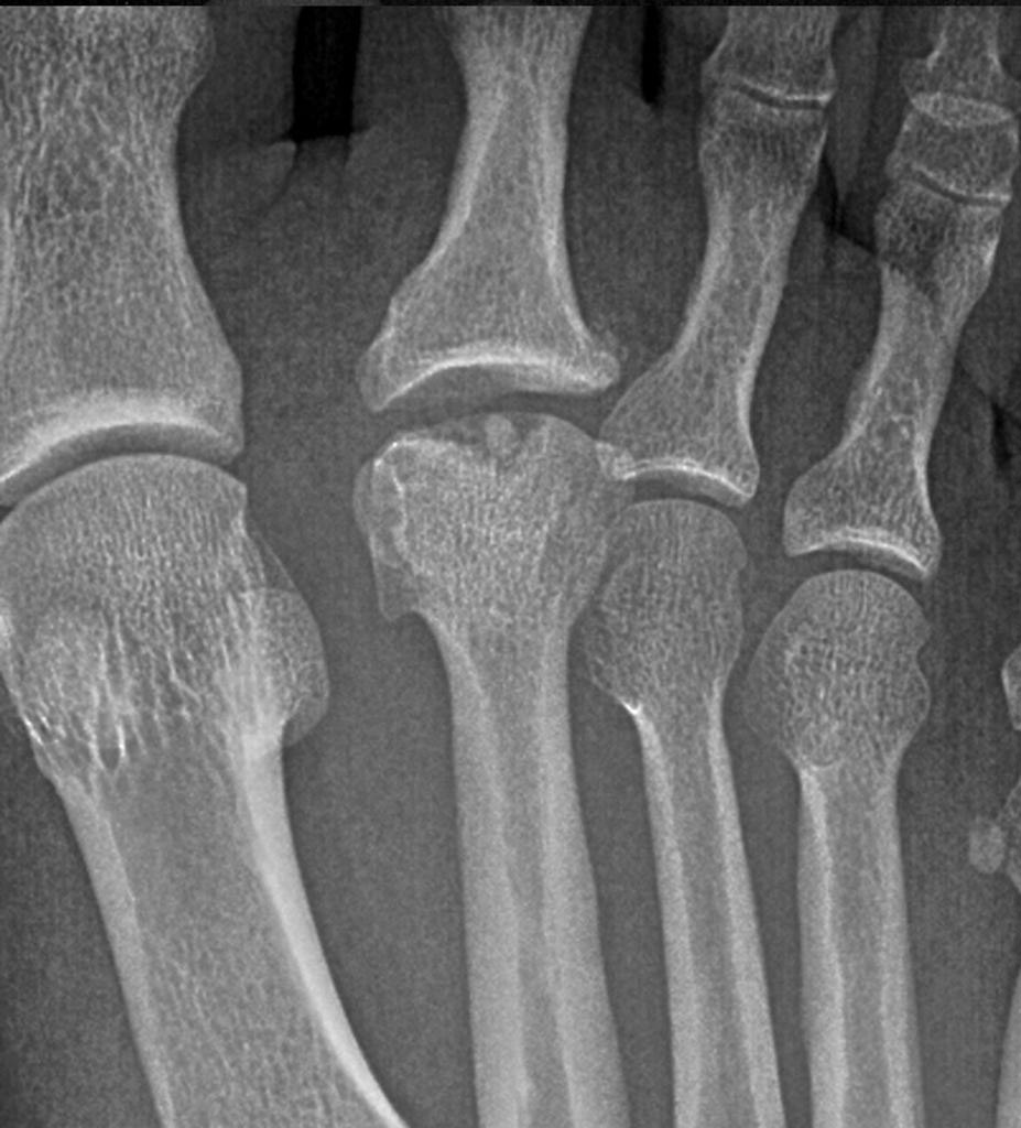
Fig 1.
MRI
MRI can show patchy edema in the metatarsal head.
Treatment
In most cases, conservative therapy is adequate. This includes rest, crutches, and plaster cast support during the acute phase of the disease.
Once the acute symptoms subside, an orthosis in the form of a metatarsal bar support can provide a degree of comfort and support.
If conservative therapy fails, a number of surgical procedures have been described that either attempt to modify the disease process or to salvage the situation.
These include:
Excision of loose bodies (Freiberg)
Elevation of the depressed metatarsal head (Smillie)
Dorsiflexion osteotomy of the metatarsal head (Gauthier)
Metatarsal shortening osteotomy (Smith)
Excision base of proximal phalanx (Trott)
Metatarsal head excision (Giannestras)
In his original series, Freiberg [1] stressed the importance of the presence of loose bodies in the MP joint. He stated that the presence of loose bodies
may be the reason for the failure of conservative treatment. He performed arthrotomies on two of the three patients with intra-articular loose bodies. There was excellent relief of symptoms.
Hoskinson [4] agreed with this approach. Trott [12], however, cautioned that it did not relieve the symptoms in all cases. Stanley et al [13] found that the presence of loose bodies was not an important predictor of the final outcome.
Smillie [8] proposed a technique of exploring the MP joint, elevating the depressed segment of the metatarsal head, and buttressing the repair
with cancellous bone graft. This procedure was indicated so long as the depressed fragment remained attached with a rim of hyaline cartilage. His results demonstrated almost perfect restoration of the articular surface. He, however, made no comments about the functional outcome of his cases.
Gauthier [3] also recommended a similar philosophy of reconstructing the metatarsal head. Based on his observation that the lesion was situated on the plantar aspect of the metatarsal head, he performed a dorsiflexion osteotomy of the metatarsal head and excised the abnormal segment. He obtained good results in 52 of his 53 cases. The procedure can be technically difficult and may further damage the already compromised metatarsal head.
Surgery is usually offered to those patients who have no lasting benefit from a 6-month trial of conservative therapy. A shortening osteotomy of the metatarsal is usually performed by excising approximately 4 mm of the metatarsal neck. The osteotomy is stabilized with a small T-shaped plate and the patient is immobilized in a below-knee plaster cast for 4 weeks. The rationale for this approach is that it decompresses the joint allowing repair of the subchondral bone. If the disease has progressed a lot then such reconstructive procedures are inappropriate. In such situations, Trott's [12] technique of partial resection of the proximal phalanx with syndactylization of the second to the third toe may be appropriate. Trott claims that the procedure provides relief of symptoms and a reasonable cosmetic outcome.
Giannestras [14] was of the opinion that metatarsal head resection was appropriate for severe disease. Hoskinson [4] reported unsatisfactory results with this procedure.
Prognosis
Not much is known of the long-term outcome of treatment of patients with Freiberg's disease, irrespective of the method of treatment used. Symptoms usually resolve with time, hence patients are rarely seen in orthopedic clinics. Hoskinson's [4] had the longest series with 28 patients who were reviewed at a mean of 12 years following diagnosis. Of the 16 patients who were treated conservatively, 11 remained asymptomatic with only 2 patients complaining of persistent aches. Twelve patients were treated by a variety of surgical methods. Eight of them continued to complain of troublesome residual symptoms.
There is still a lot to learn about the etiology and treatment of this condition. Since the condition is relatively rare, different forms of treatment have never been subjected to a prospective randomized trial.
References
Freiberg AH: Infraction of the second metatarsal bone. A typical Injury. Surg Gynecol Obstet 19:191, 1914.
Wagner A: Isolated aseptic necrosis in the epiphysis of the first metatarsal bone. Acta Radiol 11:80, 1930.
Gauthier G. Elbaz R: Freiberg's infraction: a subchondral bone fracture. A new surgical treatment. Clin Orthop 142:93, 1979.
Hoskinson J: Freiberg's disease: A review of the long-term results. Proc R Soc Med 67:10, 1974.
Crock HV: The Blood Supply of the Lower Limb Bones in Man. Livingstone Ltd., Edinburgh and London, 1967.
Freiberg AH: The so-called infraction of the second metatarsal bone. J Bone Joint Surg 8:257, 1926.
Kohler A: Grenzen des Normalen und Anfange des Pathologischen. p. 77. In Rontgenbilde. LucasGrafe and Sillem. Hamburg. 1924.
Smillie IS: Freiberg's infraction (Kohler's second disease). J Bone Joint Surg 39B:580, 1957
Braddock GTF: Experimental epiphyseal injury and Freiberg's disease. J Bone Joint Surg 4lB:154, 1959.
Stanley D, Belts RP, Rowley DI, Smith TWD: Assessment of the aetiological factors in the development of Freiberg's disease. J Foot Surg 29:444, 1990.
Belts RP, Stanley D. Smith TWD: Foot pressure studies in Freiberg's disease. Foot 1:21, 1991.
Trott AW: Developmental disorders. In Jahss MH (ed): Disorders of the Foot, WB Saunders, Philadelphia, 1982.
Stanley D, Smith TWD, Rowley DI: The conservative and surgical management of Freiberg's disease. Foot 1:97, 1991.
Giannestras NJ: Foot Disorders: Medical and Surgical Management. 2nd Ed. Lea & Febiger, Philadelphia, 1973.
No comments:
Post a Comment