Necrotizing Fasciitis
DR KS Dhillon
Introduction
In 1871 Jones first described necrotizing skin infections. At the time the term hospital gangrene was generally used (1). Wilson coined the term necrotizing fasciitis (NF) in the 1950s to describe necrosis of the fascia and subcutaneous tissue with relative sparing of the underlying muscle (2). In patients with necrotizing fasciitis there is rapid destruction of tissue, systemic toxicity, and, if not treated aggressively, gross morbidity and mortality. Early diagnosis and aggressive treatment reduces the risk. It is, however, often difficult to diagnose NF. Sometimes patients are treated for cellulitis until there is rapid deterioration (3). Antibiotic therapy with early surgical exploration and debridement is necessary for a good outcome.
Classification
There have been different classifications and terms used to describe necrotizing infections of the skin and subcutaneous tissue. Some of these include synergistic necrotizing cellulitis, streptococcal myonecrosis, necrotizing fasciitis, and gas gangrene. These classifications and terminology have been based on affected anatomy, depth of infection, and microbial cause.
Terms such as myonecrosis, necrotizing fasciitis, and necrotizing adipositis refer to classification by depth of infection. Type 1 and type 2 infections are based on microbial cause. Necrotizing infections are classified according to anatomical sites. Fournier gangrene for example involves the perineum and Ludwig angina involves the submandibular and sublingual spaces. These infections are named after the physicians who initially described them (4,5).
These descriptive terms can cause much confusion. Anaya et al (6) recently proposed that the term necrotizing soft tissue infections should be used to describe them all, as their treatment with early surgery and broad-spectrum antibiotics is the same.
Microbiology
NF has been classified microbiologically as either type 1 which is polymicrobial or type 2 which is monomicrobial (7). Polymicrobial infections are generally more common with a mixture of aerobic and anaerobic organisms (8). Polymicrobial infections typically occur in the trunk and perineum. These microbes reflect normal skin commensalism found adjacent to the site of infection. In NF of the perineum, anaerobic bacteria are isolated. The isolates consist of Gram-positive organisms, such as S pyogenes, staphylococcus aureus, and enterococci. Gram-negative aerobes, such as Escherichia coli and Pseudomonas species; and anaerobic organisms, such as Bacteroides or Clostridium species are also found (9,10). Type 1 NF occurs in immunocompromised individuals. These include patients with diabetes mellitus or chronic renal failure.
Polymicrobial infections are more common than monomicrobial infections. These infections typically occur in the limbs and affect healthy patients with no implicative comorbidities. There is usually a history of trivial trauma. The usual pathogens are S pyogenes and S aureus. Type 2 NF might be associated with toxic shock syndrome (6). Community-acquired methicillin-resistant S aureus (MRSA) has become common in NF. A recent retrospective review by Miller et al of cases from 2000 to 2006 in Los Angeles, California, showed MRSA isolates in one-third of cases (10).
Pathology
Most cases start with trauma to the skin surface. A penetrative injury or even acupuncture needles can seed bacteria into the deeper tissues. The infection begins in the deep tissue planes, and the epidermis might not be affected initially. The clinical disease becomes obvious when the bacteria spreads through the tissue along the deep fascia. After the bacteria has penetrated the deeper tissues, they rapidly multiply within viable tissue. Fibrous attachments between subcutaneous tissues and fascias limit the spread to areas like the feet, hands, and scalp. The lack of fibrous attachments in the trunk and limbs can lead to widespread infection and tissue damage. Infection also spreads to lymphatic and venous channels. This leads to edema. The spread of bacteria can cause thrombosis of blood vessels in the dermal papilla. This can result in ischemia and gangrene of subcutaneous fat and dermis (11). If the fascia is breached, infection of the muscle occurs which can lead to myositis. The gas-producing organisms such as Clostridium species can produce subcutaneous gas leading to gas gangrene.
Infections with toxin-producing bacteria such as S aureus and S pyogenes can lead to a toxic shock-like syndrome. Seemingly limited infection can produce septic shock and multiorgan failure.
Risk factors
Patients presenting with NF usually have some predisposition to infection. Predisposing risk factors include:
Diabetes
Chronic disease
Peripheral vascular disease
Renal failure
Underlying malignancy
Immunosuppressive drugs (eg, prednisolone)
Malnutrition
Age > 60 years
Intravenous drug misuse
Obesity
A Singapore study by Wong et al showed that 70.3% of patients with NF had diabetes mellitus (12). Most patients have a history of trauma, surgery, or penetrating injury. The injury, however, can be quite trivial such as due to insect bites or scratches.
NSAID use has been implicated in severe necrotizing streptococcal infections. Nonsteroidal anti-inflammatory drugs apparently impair lymphocyte function. Anecdotal reports suggest that there is an association between the use of NSAIDs and the progression of invasive group A streptococcal infections to shock and multiorgan failure (13). However, it could also be possible that suppression of symptoms and signs of inflammation leads to a delay in diagnosis, especially in patients presenting early with nonspecific symptoms (14).
In the pediatric population, risk factors for NF include malnutrition and skin infections such as varicella (15,16).
It is important not to rule out NF in normal healthy patients with minor dermatologic trauma.
Clinical features
Patients having NF can present with constitutional symptoms of sepsis such as tachycardia, fever, altered mental state, and diabetic ketoacidosis alone or with evidence of skin inflammation. Infection commonly occurs on the limbs.
According to a retrospective review by Golger et al of patients treated for NF in 3 tertiary hospitals in Canada, the common sites of infection were the lower extremities (28%), upper extremities (27%), perineum (21%), trunk (18%), and the head and neck (5%) (17). NF often first starts in the deep tissue planes, hence at initial presentation, there might be minimal epidermal involvement. This makes it difficult to differentiate from cellulitis and non-necrotizing skin infections.
Patients with NF often have systemic toxicity. They initially present with fever which is greater than 38°C, diaphoresis, tachycardia, and even an altered mental state or diabetic ketoacidosis. Physical examination includes all parts of the body to search for inflammation of the skin. This is necessary, especially in patients who present with sepsis where the source is not obvious. The oral cavity and perineum are areas that can be easily missed (18).
Most patients with NF present with signs and symptoms of skin inflammation such as pain, erythema, and skin edema. Such signs and symptoms, however, are also present in less serious conditions such as cellulitis and erysipelas. The degree of pain relative to the skin condition might provide the physician with clues. NF usually presents with pain that is out of proportion to the degree of skin inflammation.
Erysipelas is an infection of the superficial dermis. It has well-defined borders and can blister profoundly. In patients with cellulitis, there is erythema with lymphangitis and some blistering.
NF usually presents with patchy discolouration of the skin with swelling and pain but without a defined margin or lymphangitis (11,19) Fig 1.
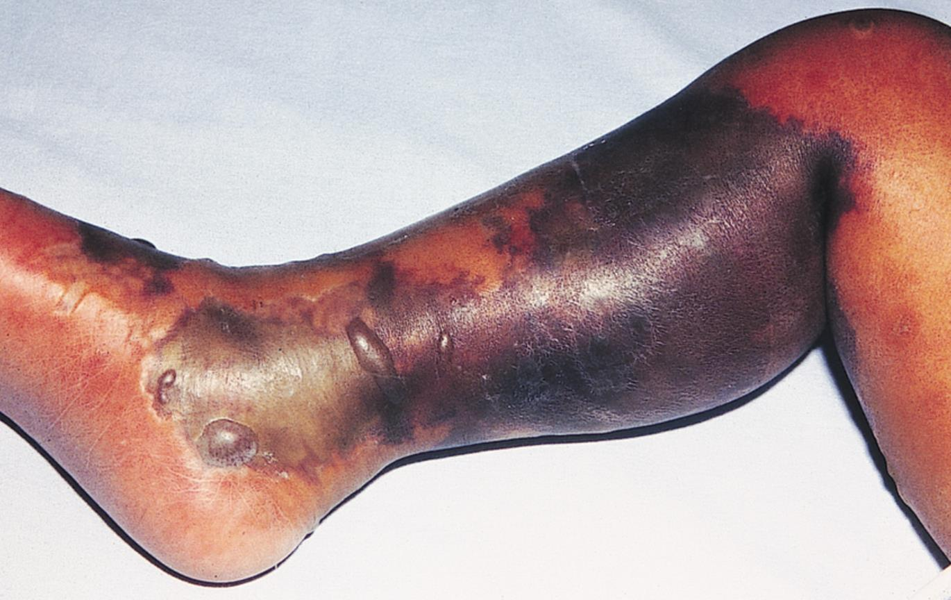
Fig 1
Progression of NF is associated with the development of tense edema, vesicles, bullae, necrosis, a grayish-brown discharge, and crepitus (20). Hemorrhagic bullae and crepitus are signs that there is likelihood of underlying fascia and muscle being compromised (21). Crepitus is a later sign that is found in only about 18% of the cases (9).
Crepitus and blistering are the most specific signs of necrotizing soft tissue infection. They are however not diagnostic. Retrospective case series, by Wang et al (13) and Elliot et al (22) showed the absence of crepitus in 62% to 63% of cases and an absence of blistering in 76% to 95% of cases on initial presentation. Lymphangitis and lymphadenopathy are absent in necrotizing infections, but they remain features of cellulitis (11,19).
Localized pain is another feature of NF. Since the disease is a deep-seated infection, the epidermis is minimally involved at first presentation. The patient may complain of pain that is out of proportion to the degree of dermal involvement or pain that extends past the margin of infection (20). The pain due to cellulitis can be treated with analgesics. The pain in patients with NF is often severe. This can make the patient exceedingly apprehensive and very fearful when examined. Certain patients like those with diabetic neuropathy with loss of sensation, can experience minimal pain leading to a missed diagnosis. This is likely in concealed sites of infection, such as the oral cavity or perineum. There can be a patch of anesthesia over the site of erythema. This is believed to be due to infarction of cutaneous nerves in necrotic subcutaneous fascia and soft tissue (23).
Disease progression
Progression of necrotizing fasciitis can follow a subacute or a hyperacute course. Patients with a hyperacute course present with sepsis and multiorgan failure. The diagnosis of sepsis is obvious, and these patients need to be hospitalized.
A subacute variation of NF has been described by several authors (24-26). Patients with this subacute variant have an indolent disease course, with festering soft tissue infection. Once the infection reaches a certain threshold, sudden deterioration occurs. Aggressive surgical debridement is needed in these cases. Progression of disease invariably occurs in this group. A delay in diagnosis can lead to greater soft tissue loss and mortality (24-26). Wong et al (25) described this entity of subacute NF as having a slow indolent course with an absence of systemic disturbance. They showed that there is gradual tissue necrosis with progressive cutaneous changes over the affected site. The progression of disease occurs despite the use of antimicrobial medications. This progression of disease leads to a sudden deterioration with rapid progression of NF or systemic features of sepsis (25).
Subacute NF can present a diagnostic dilemma hence there is a need to be aware of it.
Hospitalization
Clinical judgment is necessary to decide which patients who present with evidence of skin inflammation should be hospitalized or receive further evaluation. Studies have shown that only 15% to 34% of patients with a discharge diagnosis of necrotizing skin and subcutaneous infection had an admitting diagnosis of NF (12,27).
Some patients with non-necrotizing soft tissue infections will require hospitalization. A history of pyrexia, diabetes mellitus, hand infections, and an area of inflammation greater than 70 square cm are independent predictors of hospitalization (28).
Patients with soft tissue infection who have signs and symptoms of systemic toxicity such as hypothermia, fever, tachycardia, hypotension, an altered mental state, severe infection, intractable nausea and vomiting, immunocompromise, failure of outpatient treatment, and poor social support would require hospitalization. The Infectious Diseases Society of America skin and soft tissue infection guidelines suggest that hospitalization should be considered in patients who have hypotension and/or an elevated creatinine level, low serum bicarbonate level, elevated creatine phosphokinase level, or a C-reactive protein level of more than 13 mg/L (29).
Cutaneous manifestations are a continuum in the early stages of NF. The signs and symptoms evolve over time (3). At the first encounter, the doctor might diagnose uncomplicated cellulitis, it is however prudent to advise the patient early review if symptoms or signs progress.
Diagnosis and surgical exploration
The diagnosis of NF is confirmed by obtaining tissue for histology during surgical exploration. When surgical exploration is carried out tissue integrity and depth of invasion can be assessed. Necrosis of the fascia and loss of fascial integrity indicates necrotizing infection. Muscle involvement is seen in the advanced stage of the disease.
Laboratory and radiologic tests are sometimes useful to help decide which patients require surgical exploration, especially in those with equivocal clinical signs.
Leukocytosis with neutrophilia, acidosis, impaired renal function, raised creatinine kinase levels, altered coagulation profile, and raised inflammatory markers, such as C-reactive protein levels, are all useful if viewed within the whole clinical context.
Clinical scores such as the laboratory risk indicator for NF (LRINEC) score are available to help diagnose NF and differentiate it from other soft tissue and skin infections (11,30- 35). A score of 6 and above indicates NF. A study by Brogan et al (16) showed that an intermediate to high risk of NF had a positive predictive value of 92% and a negative predictive value of 96%. The LRINEC score was based on retrospective studies of patients with highly suspected or diagnosed NF. It has, however, not been validated in patients for whom the diagnosis of NF is not apparent in the initial assessment. Blood cultures are usually part of the workup in hospitals. The blood cultures can yield up to 27.3% positive cultures in necrotizing infections (8) compared with 2% positive blood culture yield in patients with cellulitis (36).
In patients with NF plain X-rays can show subcutaneous gas. This is however not a sensitive finding. It is positive in fewer than 25% of cases. The absence of gas does not exclude NF (37). When the diagnosis is in doubt and the signs are equivocal computed tomography (CT) scans and magnetic resonance imaging (MRI) might be useful. Important imaging findings include fat stranding, asymmetrical fascial thickening, and gas tracking along fascial planes. CT scans are estimated to have a sensitivity of 80% for detecting necrotizing soft tissue infections (38). An MRI will show subcutaneous thickening with fluid collection in patients with cellulitis. When there is deep fascia involvement with fluid collection, enhancement, and thickening, following contrast administration, necrotizing infections has to be considered (36). Schmid et al (39) in a study showed that the sensitivity of MRI is 100% with a specificity of 86% in patients with NF. This has been disputed by other authors who have stated that in early cases of NF, MRI might not show fascial involvement (40). If the clinical suspicion is high, doctors can opt to explore and perform tissue biopsies.
Bedside tests that can be carried out include needle aspiration and incision biopsy. Negative results do not, however, exclude NF. Surgical exploration should be carried out.
The diagnosis of NF is clinical, and the clinician obtains information from both the patient’s condition and the various tests done. There has to be a high index of suspicion and a low threshold for surgical referral.
Surgical exploration will show gray necrotic tissue, “dishwater” pus, noncontracting muscle, lack of bleeding, thrombosed vessels, and a positive “finger test” result. The “finger test” is characterized by a lack of resistance to finger dissection in normally adherent tissues (6).
Treatment
Once the diagnosis has been made the treatment should begin immediately on multiple fronts. Urgent surgical consultation is obtained with the intention of early wound debridement for excision of all nonviable tissue, collection of tissue cultures, and delineation of the extent of the disease. This is important as tissue hypoxia limits the efficacy of intravenous antibiotics. Patients are often incredulous when told they need an extensive operation for their skin infection. They have to be educated about the gravity of their condition and the risk of increased mortality if surgery is not performed.
Wide-spectrum intravenous antibiotics are started while waiting for the blood culture results. These antibiotics should cover S pyogenes, S aureus, Gram-negative aerobes, and anaerobes. Gram-negative organisms are likely in the abdominal wall and perineal wounds, necrotic diabetic foot ulcers, and in heavily contaminated wounds.
Hyperbaric oxygen has also been used in addition to antibiotics and surgery. The role of hyperbaric oxygen is still ill-defined. Some authors have reported a reduction in morbidity, mortality, and the need for repeated debridement in up to about two-thirds of cases (41,42). Well-controlled randomized trials are however lacking. A retrospective review by Golger et al showed that morbidity associated with NF was higher in patients who had hyperbaric oxygen therapy (17).
Intravenous immunoglobulins might play a therapeutic role in type 2 NF caused by streptococci resulting in streptococcal toxic shock syndrome. Recently, a multicentre, randomized, double-blind, placebo-controlled trial by Darenberg et al (43) evaluated the safety and efficacy of intravenous immunoglobulins in streptococcal toxic shock syndrome. Although the trial was prematurely stopped because of poor recruitment, it however showed a 3.6-fold higher mortality in the placebo group compared with the treatment group.
Prognosis
Mortality due to necrotizing fascitis can be considerable. Without surgical intervention, mortality can approach 100%. When Jones first reported necrotizing infections the mortality was 46% (1). Recent data shows a mortality rate of 16.4% for community-acquired necrotizing soft tissue infections (44) and 36.3% for postprocedural necrotizing infections (45). All the patients in these studies were managed in hospitals with intravenous antibiotics and surgical interventions. Despite the medical progress in the last 135 years, mortality is still high. In patients with streptococcal toxic shock syndrome, the mortality is higher. In diabetic patients, especially those presenting with diabetic ketoacidosis or hyperosmolar hyperglycemic nonketotic acidosis the death rates are higher and the hospital stay longer (46). A delay in performing surgery of more than 24 hours is an independent risk factor for mortality (12).
There can be considerable postoperative morbidity, especially when there is extensive debridement resulting in muscle loss. Patients usually have to undergo a period of rehabilitation to regain the function of the areas affected. Disfigurement and scaring can be substantial.
Prophylaxis
Since NF is a potentially lethal disease the question arises whether those in close contact with the patient should be given chemoprophylaxis. This is particularly so in patients with type 2 infections caused by S pyogenes since this organism is highly contagious and has been responsible for epidemics of pharyngitis, scarlet fever, and surgical wound infections. The United Kingdom and Centers for Disease Control and Prevention both affirm that antibiotic prophylaxis is not warranted given currently available evidence (47-49). It is recommended that healthcare workers inform and educate household contacts of patients with invasive group A streptococcal infections, to seek medical attention immediately if they develop any symptoms.
Conclusion
Necrotizing fasciitis is a potentially life-threatening medical emergency that encompasses a devastating and rapidly spreading destruction of soft tissue with associated systemic toxicity.
NF is a rare diagnosis with a wide spectrum of presentations. The most important challenge associated with NF is establishing an early diagnosis. Initially, the skin may not be involved, which can delay treatment in patients who may appear healthy. In the initial stages, NF may be confused with cellulitis or other superficial skin infections.
The infection can be located anywhere in the body. There can be rapid progression of the infection with hyperacute systemic deterioration of the patient.
The spread of the infection can lead to limb amputation. The risk for mortality is estimated to be 15-35%. This can however reach 100% if source control is not obtained. The rapid clinical course is related to microbial virulence. Most cases are polymicrobial with organisms such as group A beta-hemolytic Streptococcus or Streptococcus pyogenes. The most important factors in patient survival are time to diagnosis and debridement.
The definitive treatment remains early and aggressive surgical intervention. Delay in treatment often results in increased risks of morbidity and mortality.
References
Jones J United States Sanitary Commission. Surgical memoirs of the War of the Rebellion. New York, NY: Hurd and Houghton; 1871. Investigation upon the nature, causes and treatment of hospital gangrene as prevailed in the Confederate armies 1861–1865; pp. 142–580.
Wilson B. Necrotising fasciitis. Am Surg. 1952;18(4):416–31.
Wang YS, Wong CH, Tay YK. Staging of necrotising fasciitis based on the evolving cutaneous features. Int J Dermatol. 2007;46(10): 1036–41.
Fournier JA. Gangrène foudroyante de la verge. Sem Med. 1883; 3:345–8.
Wasson J, Hopkins C, Bowdler D. Did Ludwig’s angina kill Ludwig? J Laryngol Otol. 2006;120(5):363–5.
Anaya DA, Dellinger EP. Necrotizing soft tissue infection: diagnosis and management. Clin Infect Dis. 2007;44(5):705–10. Epub 2007 Jan 22.
File TM, Jr, Tan JS, DiPersio JR. Group A streptococcal necrotizing fasciitis. Diagnosing and treating the “flesh-eating bacteria syndrome” Cleve Clin J Med. 1998;65(5):241–9.
Elliot D, Kufera JA, Myers RA. The microbiology of necrotizing soft tissue infections. Am J Surg. 2000;179(5):361–6.
Sudarsky LA, Laschinger JC, Coppa GF, Spencer FC. Improved results from a standardized approach in treating patients with necrotizing fasciitis. Ann Surg. 1987;206(5):661–5.
Miller LG, Perdreau-Remington F, Rieg G, Mehdi S, Perlroth J, Bayer AS, et al. Necrotizing fasciitis caused by community-associated methicillin-resistant Staphylococcus aureus in Los Angeles. N Engl J Med. 2005;352(14):1445–53.
Seal DV. Necrotizing fasciitis. Curr Opin Infect Dis. 2001;14(2): 127–32.
Wong CH, Chang HC, Pasupathy S, Khin LW, Tan JL, Low CO. Necrotizing fasciitis: clinical presentation, microbiology, and determinants of mortality. J Bone Joint Surgery Am. 2003;85-A(8): 1454–60.
Stevens DL. Could nonsteroidal antiinflammatory drugs (NSAIDs) enhance the progression of bacterial infections to toxic shock syndrome? Clin Infect Dis. 1995;21(4):977–80.
Aronoff DM, Bloch KC. Assessing the use of nonsteroidal antiinflammatory drugs and necrotising fasciitis caused by group A streptococcus. Medicine (Baltimore) 2003;82(4):225–35.
Fustes-Morales A, Gutierrez-Castrellon P, Duran-Mckinster C, Orozco-Covarrubias L, Tamayo-Sanchez L, Ruiz-Maldonado R. Necrotizing fasciitis: report of 39 paediatric cases. Arch Dermatol. 2002;138(7):893–9.
Brogan TV, Nizet V, Waldhausen JH, Rubens CE, Clarke WR. Group A streptococcal necrotizing fasciitis complicating primary varicella: a series of fourteen patients. Pediatr Infect Dis J. 1995;14(7):588–94.
Golger A, Ching S, Goldsmith CH, Pennie RA, Bain JR. Mortality in patients with necrotizing fasciitis. Plast Reconstr Surg. 2007;119(6):1803–7.
Hill MK, Sanders CV. Necrotizing and gangrenous soft tissue infections. In: Sanders CV, Nesbitt LT Jr, editors. The skin and infection: a color atlas and text. Baltimore, MD: Lipincott, Williams & Wilkins; 1995. pp. 62–75.
Green RJ, Dafoe DC, Raffin TA. Necrotizing fasciitis. Chest. 1996;110(1):219–29.
Headley AJ. Necrotizing soft tissue infections: a primary care review. Am Fam Physician. 2003;68(2):323–8.
Hsiao CT, Lin LJ, Shiao CJ, Hsiao KY, Chen IC. Hemorrhagic bullae are not only skin deep. Am J Emerg Med. 2008;26(3):316–9.
Elliot DC, Kufera JA, Myers RA. Necrotizing soft tissue infections. Risk factors for mortality and strategies for management. Ann Surg. 1996;224(5):672–83.
Dufel S, Martino M. Simple cellulitis or a more serious infection? J Fam Pract. 2006;55(5):396–400.
Wong CH, Wang YS. The diagnosis of necrotizing fasciitis. Curr Opin Infect Dis. 2005;18(2):101–6.
Wong CH, Wang TS. What is subacute necrotising fasciitis? A proposed clinical diagnostic criteria. J Infect. 2006;52(6):415–9. Epub 2005 Oct 6.
Jarrett P, Rademaker M, Duffill M. The clinical spectrum of necrotising fasciitis. A review of 15 cases. Aust N Z J Med. 1997;27(1):29–34.
Hefny AF, Eid HO, Al-Hussona M, Idris KM, Abu-Zidan FM. Necrotizing fasciitis: a challenging diagnosis. Eur J Emerg Med. 2007;14(1):50–2.
Diercks DB, Kuppermann N, Derlet RW, Ernst AA. Derivation and validation of a model for the need of hospital admission in patients with extremity cellulitis. Acad Emerg Med. 2000;7(5):562.
Stevens DL, Bisno AL, Chambers HF, Everett ED, Dellinger P, Goldstein EJ, et al. Practice guidelines for the diagnosis and management of skin and soft-tissue infections. Clin Infect Dis. 2005;41(10):1373–406. Epub 2005 Oct 14.
Green RJ, Dafoe DC, Raffin TA. Necrotizing fasciitis. Chest. 1996;110(1):219–29.
Headley AJ. Necrotizing soft tissue infections: a primary care review. Am Fam Physician. 2003;68(2):323–8.
Hsiao CT, Lin LJ, Shiao CJ, Hsiao KY, Chen IC. Hemorrhagic bullae are not only skin deep. Am J Emerg Med. 2008;26(3):316–9.
Elliot DC, Kufera JA, Myers RA. Necrotizing soft tissue infections. Risk factors for mortality and strategies for management. Ann Surg. 1996;224(5):672–83.
Dufel S, Martino M. Simple cellulitis or a more serious infection? J Fam Pract. 2006;55(5):396–400.
Wong CH, Khin LW, Heng KS, Tan KC, Low CO. The LRINEC (laboratory risk indicator for necrotizing fasciitis) score: a tool for distinguishing necrotizing fasciitis from other soft tissue infections. Crit Care Med. 2004;32(7):1535–41.
Perl B, Gottehrer NP, Raveh D, Schlesinger Y, Rudensky B, Yinnon AM. Cost-effectiveness of blood cultures for adult patients with cellulitis. Clin Infect Dis. 1999;29(6):1483–8.
Lim YJ, Yong FC, Wong CH, Tan AB. Necrotising fasciitis and traditional medical therapy—a dangerous liaison. Ann Acad Med Singapore. 2006;35(4):270–3.
Wyoski MG, Santora TA, Shah RM, Friedman AC. Necrotizing fasciitis: CT characteristics. Radiology. 1997;203(3):859–63.
Schmid MR, Kossmann T, Duewell S. Differentiation of necrotizing fasciitis and cellulitis using MR imaging. AJR Am J Roentgenol. 1998;170(3):615–20.
Arslan A, Pierre-Jerome C, Borthne A. Necrotizing fasciitis: unreliable MRI findings in the preoperative diagnosis. Eur J Radiol. 2000;36(3):139–43.
Riseman JA, Zamboni WA, Curtis A, Graham DR, Konrad HR, Ross DS. Hyperbaric oxygen therapy for necrotizing fasciitis reduces mortality and the need for debridements. Surgery. 1990;108(5):847–50.
Jallali N, Withey S, Butler PE. Hyperbaric oxygen as adjuvant therapy in the management of necrotizing fasciitis. Am J Surg. 2005;189(4):462–6.
Darenberg J, Ihendyane N, Sjölin J, Aufwerber E, Haidl S, Follin P, et al. Intravenous immunoglobulin G therapy in streptococcal toxic shock syndrome: a European randomized, double-blind, placebo-controlled trial. Clin Infect Dis. 2003;37(3):333–40.
Frazee BW, Fee C, Lynn J, Wang R, Bostrom A, Hargis C, et al. Community-acquired necrotizing soft tissue infections: a review of 122 cases presenting to a single emergency department over 12 years. J Emerg Med. 2008;34(2):139–46. Epub 2007 Aug 29.
Miller AT, Saadai P, Greenstein A, Divino CM. Postprocedural necrotizing fasciitis: a 10-year retrospective review. Am Surg. 2008;74(5):405–9.
Oncul O, Erenoglu C, Top C, Küçükardali Y, Karabudak O, Kurt Y, et al. Necrotizing fasciitis: a life-threatening clinical disorder in uncontrolled type 2 diabetic patients. Diabetes Res Clin Pract. 2008;80(2):218–23. Epub 2008 Jan 10.
Health Protection Agency, Group A Streptococcus Working Group. Interim UK guidelines for management of close community contacts of invasive group A streptococcal disease. Commun Dis Public Health. 2004;7(4):354–61.
Smith A, Lamagni TL, Oliver I, Efstratiou A, George RC, Stuart JM. Invasive group A streptococcal disease: should close contacts routinely receive antibiotic prophylaxis? Lancet Infect Dis. 2005;5(8):494–500.
Prevention of Invasive Group A Streptococcal Infections Workshop Participants. Prevention of invasive group A streptococcal disease among household contacts of case patients and among postpartum and postsurgical patients: recommendations from the Centers for Disease Control and Prevention. Clin Infect Dis. 2002;35(8):950–9. Epub 2002 Sep 26. Erratum in: Clin Infect Dis 2003;36(2):243.
No comments:
Post a Comment