Wednesday, 20 July 2022

Radial Head Fractures

    Radial Head Fractures


                     Dr. KS Dhillon


Introduction

Radial head fractures are common intra-articular fractures of the elbow. They can be associated with an injury to the distal radioulnar joint and/or to the interosseous membrane (Essex-Lopresti).

Diagnosis is made with x-rays of the elbow. CT scans can be useful for surgical planning. 

Treatment is nonoperative for undisplaced fractures without a mechanical block to motion. For displaced fractures or fractures associated with mechanical block to motion or elbow/forearm instability, the treatment is surgical.


Etiology

The radial head fracture results from a fall on an outstretched hand with the

elbow in extension and forearm in pronation. During such a fall the most force is transmitted from the wrist to the radial head. 

The incidence of associated injuries is about 30%. These include associated soft tissue injuries and skeletal injuries. The ligamentous and interosseous injuries include:

  • Lateral collateral ligament (LCL) injury is the most common with up to 80% on MRI

  • Medial collateral ligament (MCL) injury

  • Essex-Lopresti injury-- radial head fracture with distal radioulnar joint (DRUJ) injury and interosseous membrane injury

  • Elbow fractures & dislocations-- coronoid fracture, olecranon fracture, Monteggia fracture/dislocation, and the terrible triad of posterolateral elbow dislocation, radial head fracture, and coronoid fracture

  • Carpal fractures- scaphoid fracture


Anatomy

The proximal radius consists of:

  • radial head

  • radial neck

  • radial tuberosity

  • radial shaft

The radial head is 15º offset from the neck. The anterolateral third of the radial head lacks subchondral bone and is easily fractured in this area.

The articular surface has an oval-shaped concavity that articulates with the capitellum of the humerus. The nonarticular portion of the radial head is

considered a safe zone for hardware placement.

The radiocapitellar joint is a pivot joint. Sixty percent of the load transfer across the elbow joint passes through this joint. A fracture or resection of the radial head decreases the surface area available for load transfer and decreases the stability of the elbow.

The ulnar portion of the radial head articulates with the lesser sigmoid notch of the ulna to form the proximal radial ulnar joint (PRUJ). This joint is important for pronation and supination of the forearm.

The ligaments at the elbow (fig1) include the lateral collateral ligament complex and the medial (ulnar) collateral ligament (MCL). The lateral collateral ligament complex consists of the lateral ulna collateral ligament (LUCL), the radial collateral ligament (RCL), the annular ligament, and the accessory lateral collateral ligament.  

The lateral ulnar collateral ligament arises from the lateral epicondyle and inserts onto the supinator crest of the ulna. It is the primary stabilizer to varus and external rotation (hypersupination) stress. It is a more important stabilizer near elbow extension. Its deficiency results in posterolateral rotatory instability. 

The radial collateral ligament inserts into the annular ligament. The annular ligament originates on the anterior aspect of the lesser sigmoid notch and inserts on the posterior aspect of the lesser sigmoid notch. It stabilizes the proximal radioulnar joint by maintaining the radial head in contact with the ulna. 

One-third of individuals have an accessory lateral collateral ligament (ALCL) which runs from the annular ligament to the supinator crest of the ulna. 

The medial (ulnar) collateral ligament (MCL) consists of three bundles, the

anterior bundle, posterior bundle, and transverse bundle. It is the primary stabilizer to valgus stress (radial head is second). The anterior bundle arises from the inferior margin of the medial epicondyle and inserts at the sublime tubercle of the ulnar coronoid process. It is composed of a superficial and a deep layer. The transverse bundle originates from the proximal medial olecranon and runs distally to insert just distal to the coronoid. This ligament originates and inserts on the ulna, hence it does not provide significant stability. The posterior bundle originates at the posterior aspect of the medial epicondyle of the humerus and attaches to the medial aspect of the olecranon process, and it forms the floor of the cubital tunnel.


Fig 1- Ligaments of the elbow


Biomechanics

The radial head confers two types of stability to the elbow, valgus stability and longitudinal stability. The radial head is a secondary restraint to the valgus load at the elbow. It is important if MCL is deficient.

The longitudinal stability is a restraint to the proximal migration of the radius. There are contributions from the interosseous membrane and DRUJ.

The load-sharing from the wrist to the radiocapitellar joint is dependent on the radiocapitellar surface area. A loss of longitudinal stability occurs with the Essex-Lopresti injury pattern where there is a radial head fracture, DRUJ injury, and interosseous membrane disruption. The radial head must be fixed or replaced to restore stability and prevent proximal migration of the radius and ulnocarpal impaction.


Classification

The commonly used classification is the Mason Classification which was 

modified by Hotchkiss and Broberg-Morrey:

  • Type I- Nondisplaced or minimally displaced (<2mm) fracture with no mechanical block to rotation

  • Type II- Displaced >2mm or angulated fracture with possible mechanical block to forearm rotation

  • Type III- Comminuted and displaced fracture with a mechanical block to motion

  •  Type IV-  Radial head fracture with associated elbow dislocation


The advanced classification is the OTA Classification 

  • 2R1A- Extra-articular pattern

  • 2R1B- Partial articular pattern

  • 2R1C- Complete articular pattern


Clinical Presentation

The most common symptom is pain. Examination shows tenderness along the lateral aspect of the elbow with limitation of elbow and forearm motion, particularly supination/pronation.

Inspection of the limb will show ecchymosis and swelling over the lateral aspect of the elbow. A deformity will be seen if there is elbow dislocation. A mechanical block to elbow motion must be looked for. Aspiration of joint hematoma and injection of local anesthesia helps in the evaluation of mechanical block. 

Stability testing of the elbow is carried out. The posterolateral drawer test,  posterolateral pivot shift test (tests LUCL), and valgus stress test (tests MCL) are carried out. 

For DRUJ injury, palpate the wrist for tenderness. Any translation in the sagittal plane of more than 50% compared to the contralateral side is abnormal. If difficult to determine DRUJ injury on examination, a dynamic CT scan in neutral, pronation and supination can be done.

For interosseous membrane injury palpate along the interosseous membrane for tenderness. 


Imaging

The recommended x-rays include AP and lateral view of the elbow and  

AP and lateral views of the forearm/wrist. 

A fracture with or without displacement and with or without intra-articular involvement can be seen. Anterior and or a posterior fat pad sign can be seen in patients with occult minimally displaced fracture of the radial head.  

The fat pad sign indicates the presence of intra-articular hemarthrosis. The posterior fat pad sign is more sensitive for a fracture.

A radiocapitellar view or Greenspan view (fig 2) can be useful for the diagnosis of a radial head fracture. It is an oblique lateral view of the elbow that is taken with the beam angled 45 degrees cephalad. It allows visualization of the radial head without coronoid overlap. It helps detect subtle fractures of the radial head.

A CT scan is carried out for comminuted fractures to further delineate the fracture fragments. It is also useful in patients with complex fracture dislocations. The CT scan can be helpful in planning surgical techniques and approaches.
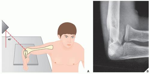

Fig 2- Radiocapitellar view or Greenspan view


Treatment


Nonoperative Treatment

Isolated minimally displaced fractures with no mechanical block (Mason Type I) are treated with short periods of immobilization for 3-7 days. Early mobilization is necessary to prevent elbow stiffness.  The outcome is generally good with good results in 85% to 95% of patients.


Operative Treatment

Open reduction and internal fixation (ORIF) is indicated in:             

  • Mason Type II fracture with mechanical block

  • Mason Type III fracture where ORIF is feasible

  • When there are other complex ipsilateral elbow injuries


The fractures can be fixed with screws alone or with a plate and screws depending on the type of fracture.

The outcome is generally good to excellent in more than 90% of the patients with Mason II fractures. The outcome of Mason III fractures is variable.

The outcome of open reduction and internal fixation is worse when there are more than 3 fragments as compared to when there are less than 3 fragments. Unsatisfactory outcome is seen in more than 50% of patients following ORIF in patients with more than 3 fragments.

There is no significant difference in outcome at 4 years in patients who had 

ORIF of isolated radial head fractures versus complex radial head fractures. Isolated fractures, however, have a better Patient-Rated Elbow Evaluation score, lower complication rate, and lower rate of secondary capsular release. 

Partial excision of the radial head or fragment excision can be carried out in

older, lower demand patients with complex fractures who have no associated instability. Fragments less than 25% of the surface area of the radial head or 25%-33% of capitellar surface area can be excised. 

Sometimes even small fragment excision can lead to instability.

Complete excision of the radial head is carried out in low demand, sedentary patients. In a delayed setting complete excision of the radial head is carried out for continued pain due to an isolated radial head fracture.       

Complete excision of the radial head is contraindicated in patients with 

destabilizing injuries such as forearm interosseous ligament injury (>3mm translation with radius pull test), coronoid fracture, and medial collateral deficiency.

The outcome after radial head excision is worse with regard to strength, function, and motion when compared to open reduction and internal fixation. The incidence of osteoarthritis after excision of the radial head is as high as 73% when compared to the contralateral uninjured elbow. 

Radial head arthroplasty is indicated in patients with:

  • Comminuted fractures (Mason Type III) with more than 3 fragments

  • Severe plastic deformity of the radial head

  • Nonunion/malunion

  • Elbow fracture-dislocations with terrible triad or Monteggia variants and with involvement of more than 30% of the articular surface of the radial head

  • Essex-Lopresti lesions- radial head excision will exacerbate elbow/wrist instability and may result in proximal radial migration and ulnocarpal impingement

Radial head fractures that require replacement have shown good clinical outcomes with metallic implants. Compared to ORIF for fracture dislocations and Mason Type III fractures, arthroplasty provides greater stability, lower complication rate, and higher patient satisfaction.


Indications and outcomes of radial head excision

The most common indication for primary excision of the radial head is acute comminuted fracture of the radial head. In the chronic setting rheumatoid arthritis is a common indication.

The literature comparing radial head excision to ORIF in the acute setting is less conclusive. There are two studies in the literature. One study found no significant differences, and one found significant differences favoring ORIF only in certain outcome measures such as grip strength and forearm rotation. Hence, there is not enough data to support one procedure over the other, even though from a clinical point of view ORIF is typically performed for those fractures involving less than three fragments that are amenable to fixation [1]. In the chronic setting also the conclusions are unclear. 

The results of secondary excision of the radial head following trauma showed a relatively high rate of post-operative pain, low satisfaction, and a high rate of giving up sports and jobs. This, however, was supported only by two studies. One study comparing primary excision within three weeks to secondary excision found that the best results were obtained after primary excision [2]. 

Most of the published studies conclude that radial head excision results in favorable long-term outcomes when used to treat isolated displaced and comminuted radial head fractures. It is important to evaluate the elbow for associated injuries prior to resection of the radial head, particularly ligamentous injuries because the outcomes in this group has been reported to be poor [3-7]. Radial head excision is contraindicated in patients with MCL or interosseous membrane injuries unless these injuries are concurrently treated.

Several studies have shown that the long-term (more than 14 years) outcome of radial head excision was good and that it was a viable treatment option with good functional results [3,6,8,9,10,11].

Radial head excision does have some long-term complications. These include osteoarthritis of the elbow, valgus instability, stiffness, and proximal migration of the radius. 

Advanced osteoarthritic changes are often present following radial head excision but typically these radiographic findings are not associated with functional impairment [9,12]. The degenerative grading does not necessarily correlate with clinical outcome [13].

Some long-term follow-up studies demonstrate valgus instability, stiffness, or proximal migration of the radius following radial head excision [14,15]. Excision of the radial head leads to a very low complication rate, with low-clinical-impact radiographic arthritis being the most significant one. There is not much literature comparing the outcome of radial head excision to radial head replacement for acute treatment of the radial head fracture. There is only one study that compared radial head excision to replacement. The study found no difference between the two treatments in terms of functional scoring but did find that extension was significantly more restricted in the replacement group [16].

There are several studies in the literature that assessed outcomes of radial head replacement for fracture with a mean follow-up of eight years or more. All the studies reported sustained good or excellent clinical results with few complications [17-20]. The contention of whether radial head replacement or excision is a better treatment for acute trauma cases remains a grey area, except where excision is contraindicated such as in situations when valgus or axial instability is present [21].

In conclusion, the primary indication for radial head excision is fracture comminution in the acute setting. The outcome is generally good or excellent following radial head excision for radial head fractures. There is insufficient evidence to recommend ORIF or replacement compared to radial head excision in patients with complex radial head fractures. Radial head excision should not be performed when there are concurrent ligamentous injuries of the elbow. Secondary radial head excision in posttraumatic situations produces inferior results and patients often continued to have residual pain. 


Treatment Techniques


1. Nonoperative management

An arm sling or posterior long arm splint can be used for 3-7 days followed by early mobilization of the elbow to prevent stiffness.


2. Open reduction and internal fixation (ORIF)

Approaches

There are several approaches for open reduction and internal fixation of the fracture. These include:

  • Kocher approach (fig 3) - In this approach, the radial head is exposed through the interval between the extensor carpi ulnaris and the anconeus. The posterior interosseous nerve (PIN) and the radial nerve must be protected. The posterior fibers of the supinator are incised and the capsule is incised in the mid-radiocapitellar plane anterior to crista supinatoris to avoid damaging the lateral ulna collateral ligament (LUCL). The risk of PIN injury is less than with the Kaplan approach (more posterior). The PIN crosses the proximal radius from anterior to posterior within the supinator muscle 4 cm distal to the radial head. In both Kocher and Kaplan's approach, the forearm should be pronated to protect PIN. The PIN originates approximately 1.2mm from the radiocapitellar joint. Pronation of the forearm pulls the nerve anteriorly and away from the surgical field. There is a risk of destabilizing the elbow if the capsule incision is too posterior and LUCL is cut, which lies below the equator of the capitellum

  • Kaplan approach (fig 3) - In this approach, the radial head is exposed through the interval between the extensor digitorum communis and extensor carpiradialis bravis. The mid-fibers of the supinator are incised and the capsule is incised anterior to the mid-radiocapitellar plane. There is less risk of disrupting LUCL and destabilizing the elbow than with the Kocher approach. There is improved exposure of the anterior fractured fragments when screw fixation is performed. There, however, is a greater risk of PIN and radial nerve injury and the approach is less extensile.
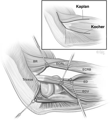


Fig 3- Kaplan and Kocher approach


  • Extensor digitorum communis (EDC) split - The incision is made longitudinally through the middle of the EDC to the origin on the lateral epicondyle. It provides improved access to the anterior half of the radial head. It also reduces the risk of iatrogenic injury to the lateral collateral ligament complex.

  • Posterior approach - There is no true intermuscular interval at the posterior aspect of the elbow. A large lateral skin flap is raised to approach the posterior aspect of the elbow. This approach is usually used when there is an associated olecranon or Monteggia fracture. This approach allows access to both the medial and lateral sides of the elbow. This approach is not so popular due to skin flap-related complications.


Technique  

  1. Screw(s) - Screws are utilized alone in simple partial articular fractures of the radial head. Herberts headless screws are used to fix articular fracture fragments. Fixation with screws produces better elbow range of motion and functional outcome scores at 1 year compared to plate fixation.

  2. Plates and screws - Plates and screws are used for fractures involving the head and neck of the radius. Mini-fragment (1.5 - 2.0 mm) plates and screws are used to fix the fracture. The plate is placed posterolaterally on the non-articular safe zone that consists of a 90-110º arc defined by the projections of the radial styloid and Lister's tubercle. The bicipital tuberosity is the distal limit of plate placement. Anything distal to that will endanger the PIN. Implants on the articular surface have to be countersunk. Plate removal is relatively common in order to restore forearm rotation. 

Complications

Several complications can result from ORIF of radial head fractures. Some of these include:

  • PIN injury

  • Destabilization of the lateral ligament complex

  • Articular surface penetration with screws 

  • Mechanical block to motion by hardware


3. Fragment Excision

If the fracture is less than 25% of the surface area of the radial head and does not compromise elbow stability, the fragment can be excised. A Kocher or Kaplan approach is used for the excision. If the fragment is too large excision can lead to elbow instability. 


4. Radial Head Resection

Radial head excision is carried out through the Kocher or Kaplan approach. 

Enough radial head should be removed to fully remove comminuted fragments of the radial head and the annular ligament must be kept intact.

Complications following radial head resection include muscle weakness, wrist pain, valgus elbow instability, heterotopic ossification, elbow arthritis, 

proximal radial migration, decreased strength, and cubitus valgus.


5. Radial head arthroplasty   

There are several types of prostheses for radial head arthroplasty. These include:


1. Metal prostheses - There are 3 types of metal prosthesis:

  • Loose stemmed prosthesis - They act as a stiff spacer. They are believed to "settle in" to anatomic position throughout the arc of motion.

  • Press-fit prosthesis - They depend on osteointegration and tight canal fit. They are prone to incorrect intramedullary positioning given their tight fit.

  • Bipolar prosthesis - They have an articulation at the head-neck junction. They allow better articulation of the radial head to the capitellum throughout the arc of motion. They may cause elbow instability when the radial head angles relative to the radial stem.


2. Pyrocarbon prostheses - Pyrocarbon implants approximate the modulus of cartilage and hence reduce the risk of capitellar wear from the metallic radial head implant. These implants are currently still under investigation. 


3. Silicon replacements (Sylastic)- These are no longer used. These replacements are independent risk factor for revision surgery due to

implant fracture and reactive synovitis.


There are 2 types of implant design namely:

  • Monoblock design where the head and stem are a single connected piece implanted together.

  • Modular design where the head and stem are 2 distinct pieces that are attached during implantation. 


Complications of radial head arthroplasty include: 

  • Radiocapitular overstuffing.  Radiocapitular overstuffing can lead to capitellar wear problems and malalignment instability. When the length of the prosthesis is excessive it produces abnormal loads on the capitellum and this produces pain, stiffness, and progressive capitellar erosion. When the diameter of the radial head prosthesis is not correct there is a cam effect that produces abnormal loads through the lateral aspect of the trochlea and the lesser sigmoid notch. Overstuffing is best assessed by direct visualization under fluoroscopy. The proximal part of the implant should align with proximal lesser sigmoid notch and the deepest point of the radial head dish should be at the same level as the lateral coronoid facet. The range of motion should be assessed in flexion and extension and it should be smooth. The radial head should remain properly aligned with the capitellum with elbow extension and flexion as well as with forearm supination and pronation.

  • Loosening. Loosening is more likely with a press-fit prosthesis. In such situations, there is a higher potential for revision surgery. To prevent loosening the implant may be fixed with bone cement if needed.

  • Implant dissociation. Implant dissociation can occur with bipolar prosthesis. Metallosis and pain can result when there is a defective locking mechanism of the head on the stem.


Complications from surgical treatment of radial head fractures

1. Surgical Site Infection

The treatment for surgical site infection is incision and drainage. If there is osteomyelitis radial head excision may be necessary. Sometimes the hardware/implant may need removal when infection complicates ORIF or radial head replacement. Intravenous antibiotics are often needed for six weeks followed by oral antibiotics if the hardware/prosthesis is retained.

2. Secondary displacement of the fracture

Secondary displacement occurs in less than 5% of fractures that are initially treated nonoperatively. In such situations, operative fixation may be  necessary.

3. Posterior interosseous nerve injury (with operative management)

The risk factors for PIN injury are dissection distal to biceps tuberosity during surgery and overaggressive retraction at the radial neck. 

If neuropraxia is suspected, conservative treatment, consisting of a cock-up wrist splint, is carried out. If no recovery occurs after several months of conservative management then an EMG is done. If the nerve palsy is permanent a tendon transfer may be needed.

4. Elbow stiffness & loss of forearm rotation

The incidence of elbow stiffness and loss of forearm movements is between 3% to 20%. The risk factors are prolonged immobilization, intra-articular fracture, malunion, nonunion and heterotopic ossification.

The first line of treatment is non-operative and it includes supervised exercise therapy with static or dynamic progressive elbow splinting over a 6 month period. The primary aim is to achieve "functional" elbow range of motion i.e 100º flexion arc (30º-130º) and 100º of rotation (50º pronation & 50º supination). Sometimes operative treatment is required. There may be a need for release of contracture with or without radial head removal or replacement. Sometimes an anconeus or Achilles allograft interposition arthroplasty is necessary. 

5. Radiocapitellar joint arthritis

The risk factors for radiocapitellar arthritis are a fracture with intra-articular displacement and the use of a metallic radial head replacement. The chances of developing arthritis increases with overstuffing.

Nonoperative treatment includes activity modification, anti-inflammatories

medications and injections. Operative treatment includes radial head resection, anconeus or Achilles allograft interposition arthroplasty, and radiocapitellar hemiarthroplasty.

6. Heterotopic ossification (HO)

The risk factors for heterotopic ossification include CNS injury, burns and 

elbow fracture/dislocation with significant soft tissue injury. Six weeks of  indomethacin can be used to minimize risk after fracture/dislocation. The use of post-operative radiation for prevention and treatment is controversial. Operative removal can be considered after HO is mature. Most patients have satisfactory outcome after excision despite the presence of residual flexion contracture. About 10% of the patients can have a recurrence.  

7. Loss of hardware fixation

Loss of hardware fixation can be treated by revision fixation, radial head replacement, or radial head removal.


References

  1. Ring D, Quintero J, Jupiter JB. Open reduction and internal fixation of fractures of the radial head. J Bone Joint Surg Am 2002; 84-A: 1811–1815.

  2. Hildebrand AH, Zhang B, Horner NS, King G, Khan M, Alolabi B. Indications and outcomes of radial head excision: A systematic review. Shoulder Elbow. 2020;12(3):193-202. doi:10.1177/1758573219864305. 

  3. Morrey BF, Chao EY, Hui FC. Biomechanical study of the elbow following excision of the radial head. J Bone Joint Surg Am 1979; 61: 63–68.

  4. Fuchs S, Chylarecki C. Do functional deficits result from radial head resection? J Shoulder Elbow Surg 1999; 8: 247–251.

  5. Boulas HJ, Morrey BF. Biomechanical evaluation of the elbow following radial head fracture. Comparison of open reduction and internal fixation vs. excision, silastic replacement, and non-operative management. Chir Main 1998; 17: 314–320.

  6. Karlsson MK, Herbertsson P, Nordqvist A, et al. Long-term outcome of displaced radial neck fractures in adulthood: 16–21 year follow-up of 5 patients treated with radial head excision. Acta Orthop 2009; 80: 368–370. 

  7. Postacchini F, Morace GB. Radial head fracture treated by resection. Long-term results. Ital J Orthop Traumatol 1992; 18: 323–330.

  8. Herbertsson P, Hasserius R, Josefsson PO, et al. Mason type IV fractures of the elbow: a 14- to 46-year follow-up study. J Bone Joint Surg Br 2009; 91: 1499–1504.

  9. Iftimie PP, Calmet Garcia J, de Loyola Garcia Forcada I, et al. Resection arthroplasty for radial head fractures: long-term follow-up. J Shoulder Elbow Surg 2011; 20: 45–50.

  10. Coleman DA, Blair WF, Shurr D. Resection of the radial head for fracture of the radial head. Long-term follow-up of seventeen cases. J Bone Joint Surg Am 1987; 69: 385–392.

  11. Faldini C, Nanni M, Leonetti D, et al. Early radial head excision for displaced and comminuted radial head fractures: considerations and concerns at long-term follow-up. J Orthop Trauma 2012; 26: 236–240.

  12. Yalcinkaya M, Bagatur AE, Erdogan S, et al. Resection arthroplasty for Mason type III radial head fractures yield good clinical but poor radiological results in the long term. Orthopedics 2013; 36: e1358–e1364.

  13. Rymaszewski LA, Mackay I, Amis AA, et al. Long-term effects of excision of the radial head in rheumatoid arthritis. J Bone Joint Surg Br 1984; 66: 109–113.

  14. Ikeda M, Sugiyama K, Kang C, et al. Comminuted fractures of the radial head. Comparison of resection and internal fixation. J Bone Joint Surg Am 2005; 87: 76–84. 

  15. Ikeda M, Oka Y. Function after early radial head resection for fracture: a retrospective evaluation of 15 patients followed for 3–18 years. Acta Orthop Scand 2000; 71: 191–194.

  16. Ünlü MC, Güven MF, Arslan L, et al. Comparison of the functional results of radial head resection and prosthesis for irreparable mason type-III fracture. Ulus Travma Acil Cerrahi Derg 2018; 24: 359–363.

  17. Burkhart KJ, Mattyasovszky SG, Runkel M, et al. Mid- to long-term results after bipolar radial head arthroplasty. J Shoulder Elbow Surg 2010; 19: 965–972. 

  18. Harrington IJ, Sekyi-Otu A, Barrington TW, et al. The functional outcome with metallic radial head implants in the treatment of unstable elbow fractures: a long-term review. J Trauma 2001; 50: 46–52. 

  19. Marsh JP, Grewal R, Faber KJ, et al. Radial head fractures treated with modular metallic radial head replacement: outcomes at a mean follow-up of eight years. J Bone Joint Surg Am 2016; 98: 527–535.

  20. Sershon RA, Luchetti TJ, Cohen MS, et al. Radial head replacement with a bipolar system: an average 10-year follow-up. J Shoulder Elbow Surg 2018; 27: e38–e44.

  21. Janssen RP, Vegter J. Resection of the radial head after Mason type-III fractures of the elbow: follow-up at 16 to 30 years. J Bone Joint Surg Br 1998; 80: 231–233.

Tuesday, 28 June 2022

Modalities for Treatment of Pain

       Modalities for Treatment of Pain


                                        DR KS Dhillon


The International Association for the Study of Pain (IASP) has defined pain as “an unpleasant sensory and emotional experience associated with actual or potential tissue damage or described in terms of such damage” [1]. There are two broad categories of pain;

  • Somatic Pain: This result from the activation of nociceptors that are sensitive to noxious stimuli in cutaneous or deep tissues. The pain experienced is described as constant, aching, and gnawing. 
  • Visceral Pain: This is also mediated by nociceptors. The pain is described as deep, aching, and colicky. It is poorly localized and often referred to cutaneous sites, which may be tender. 


The five most common types of pain include:

  • Acute pain
  • Chronic pain
  • Neuropathic pain
  • Nociceptive pain
  • Radicular pain


Acute pain

Acute pain is a sudden, sharp pain that lasts less than 3 months. Acute pain tends to be related to an injury or a temporary illness. It typically subsides after the injury heals or the illness subsides. Acute pain from an injury can evolve into chronic pain if the injury doesn’t heal properly or if the pain signals malfunction.

Chronic pain

Chronic pain is ongoing pain that lasts longer than 3 months. This pain is considered a disease state and affects 1 out of 5 adults. Chronic pain is difficult to diagnose and can be misdiagnosed. Chronic pain is caused by an underlying issue. Some of the conditions that produce chronic pain include headaches, arthritis, cancer, nerve pain, back pain, and fibromyalgia. Chronic pain is longer in duration and can be constant or intermittent. 


Neuropathic pain

Neuropathic pain results from damage to nerves or other parts of the nervous system. The pain is shooting, stabbing, or burning in nature. Sometimes it feels like pins and needles. It can affect sensitivity to touch and it makes individuals have difficulty feeling hot or cold sensations. Neuropathic pain is a common type of chronic pain. It can be intermittent and it can also be so severe that it makes performing daily tasks difficult.


Nociceptive pain

Nociceptive pain is a type of pain caused by damage to body tissue that is caused by injury. People often describe it as being a sharp, achy, or throbbing pain. This type of pain is often experienced in the muscles, joints, skin, tendons, and bones. Such pain can be both acute and chronic.


Radicular pain

Radicular pain is a very specific type of pain that occurs when the spinal nerve gets compressed or inflamed. The pain radiates along the distribution of the nerve or nerve root. It can radiate from the back to the lower limbs or from the neck to the upper limbs. Individuals with radicular pain can experience tingling, numbness, and muscle weakness. 


Pain scores 

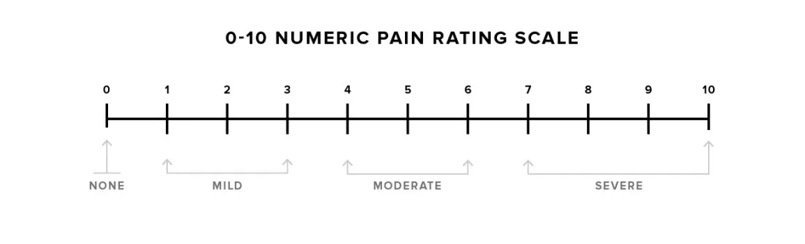
The unidimensional pain intensity scales commonly used are:

Numeric Rating Scale (NRS), 

Visual Analog Scale (VAS),

Verbal Rating / Descriptor Scale (VRS/VDS).

The VAS is the most widely used tool for estimating both severity of pain and to judge the extent of pain. The patient is asked to select a point on a line drawn between two ends to express how intense he/she perceives pain. The VAS is a continuous scale comprised of a horizontal or vertical line, usually 100 mm long, anchored by two verbal descriptors i.e., “no pain” and “worst imaginable pain”-Fig 1.


Figure 1


Pain management

Pain management should be carried out using a multimodal approach. It should consist of treatments from one or more clinical disciplines that are incorporated

into an overall treatment plan. This allows for different approaches to address the pain problem. Multidisciplinary approaches address different aspects of pain including psychosocial effects on the patient [2,3,4]. Such a coordinated, integrated approach reduces pain severity, improves mood and overall quality of life, and improves function [5,6].

Opioid and nonopioid analgesics are the main drugs used to treat pain. Antiseizure, antidepressants and other central nervous system (CNS)–active drugs are also used for chronic or neuropathic pain and can be first-line therapy for some conditions. Nerve stimulation, neuraxial infusion, and neural blockade can help in some patients.

Cognitive-behavioral interventions can reduce pain and pain-related disability and help patients cope with the pain. These interventions include counseling the patient to develop personal coping strategies and counseling the patients and their families to work together to manage pain.


Nonopioid Analgesics

Nonsteroidal anti-inflammatory drugs (NSAIDs) and acetaminophen are effective for mild to moderate pain. Most of the NSAIDs are given orally. Ibuprofen, ketorolac, diclofenac, and acetaminophen can be given parenterally. The advantage of using nonopioids is that they do not cause physical dependence or tolerance.

Acetaminophen has no antiplatelet effects and no anti-inflammatory effects and does not cause gastric irritation.

Aspirin is the cheapest NSAID, but unfortunately, it has irreversible antiplatelet effects and it increases the risk of gastrointestinal (GI) bleeding.

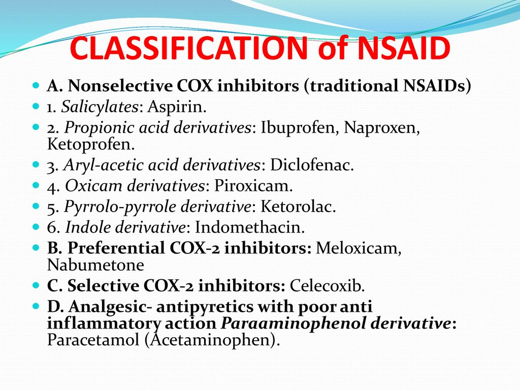
NSAIDs have analgesic and anti-inflammatory effects as well as antiplatelet effects. They inhibit cyclooxygenase (COX) enzymes and hence reduce the production of prostaglandins. There are several classes of NSAIDs, which have different mechanisms and adverse effects (Fig 1):


Fig 1.

There are 2 types of COX inhibitors i.e nonselective COX inhibitors (eg, ibuprofen, naproxen) and selective COX-2 inhibitors (coxibs; eg, celecoxib)

Both the COX inhibitors are effective analgesics. The coxibs have the lowest risk of gastric ulceration and gastritis. 

COX-2 inhibition has a prothrombotic effect that can increase the risk of myocardial infarction, claudication, and stroke. This effect varies with the type of drug, as well as by the dose and duration of use. The risk is very low with some nonselective COX inhibitors such as ibuprofen, naproxen, and coxibs. All NSAIDs should be used cautiously in patients with clinically significant atherosclerosis or multiple cardiovascular risk factors because of the potential for prothrombotic effects of all NSAIDs.  The use of NSAIDs can be associated with renal insufficiency, hypertension, and cardiac-related events.

There are no significant side effects if NSAIDs are used for a short duration. Whenever therapy is likely to be long-term (eg, months) some clinicians use a coxib first because the risk of GI adverse effects is lower. Coxib is used in patients who are predisposed to GI adverse effects, such as older patients, patients taking steroids, and those with a history of peptic ulcer disease or gastritis. 

All NSAIDs should be used cautiously in patients with renal insufficiency. 

If a lower dose provides inadequate analgesia, a higher dose is given, up to the conventional safe maximum dose. If with higher dose analgesia remains inadequate, the drug should be stopped. During long-term NSAID therapy, it is prudent to monitor for occult blood in stool and changes in the blood count, electrolytes, and hepatic and renal function.

Acetaminophen can be effective for the treatment of mild to moderate pain. The use of acetaminophen can be associated with dose-dependent liver toxicity, especially if the drug is taken at high doses, with alcohol, or by those with liver disease [7].


Opioid Analgesics

Opioid is a term used for natural or synthetic substances that bind to specific opioid receptors in the central nervous system (CNS). Opioids are also called narcotics. Opioids have both sleep-inducing and analgesic effects.

Some opioids have both agonist and antagonist actions. The potential for abuse among individuals with a known history of abuse or addiction is lower with agonist-antagonists such as buprenorphine and butorphanol than with pure agonists such as morphine, oxycodone, and hydromorphone. Agonist-antagonist drugs have a ceiling effect for analgesia and they induce a withdrawal syndrome in individuals already physically dependent on opioids.

Opioid analgesics have proven efficacy in the treatment of pain. The risk of opioid misuse has to be kept in mind when using opioids for the treatment of pain. Underuse of opioids can result in needless pain and suffering. 

Some of the reasons for underusage leading to undertreatment include:

  • Underestimation of the effective dose
  • Overestimation of the risk of adverse effects

Simultaneous treatment of the condition causing the pain can limit the duration of severe pain and the need for opioids.

For acute pain, short-acting (immediate-release) pure agonist drugs are used at the lowest effective dosage and for a short time. The Centers for Disease Control and Prevention (CDC) guidelines recommend 3 to 7 days[8]. Using opioids at higher doses and for a longer time increases the risk of needing long-term opioid therapy, adverse effects, and misuse of opioids. Opioids should not be withheld when treating cancer pain. In such cases, adverse effects can be prevented or managed, and addiction is not much of a concern.

There is no good evidence to support the use of opioids for the long-term treatment of chronic pain due to non-terminal disorders. Long-term opioid therapy can result in adverse effects such as addiction, overdose, respiratory depression, and death. In patients with chronic pain due to non-terminal disorders, lower-risk nonopioid therapies should be tried before opioids.

If nonopioid therapy has been unsuccessful in patients with chronic pain due to non-terminal disorders, opioid therapy can be considered. In such cases, opioids are used in combination with nonopioid therapies. The goals, expectations, and risks of treatment should be explained to the patient. The patient should also be counseled about the misuse of the drug.

When treatment with opioids is appropriate the chronic pain can be treated with long-acting formulations. Long-acting formulations should be avoided in opioid-naive patients because of a higher risk of serious adverse effects. 

Patients on long-term (more than 3 months) opioid therapy are regularly assessed for pain control, functional improvement, adverse effects, and signs of misuse of the drug. 

Opioid therapy should be tapered and stopped if the following occur:

  • Patients continue to have persistent severe pain despite increasing opioid doses.
  • Patients do not adhere to the treatment protocol
  • Physical or mental function does not improve.


All patients treated with opioids for more than a few days develop physical dependence i.e they develop withdrawal symptoms when a drug is stopped. They also develop tolerance i.e they develop decreased response to the same dose of a drug that is used repeatedly. Therefore, opioids should be used for as short periods as possible. In patients who have developed dependence, the dose should gradually be tapered to control withdrawal symptoms when opioids are no longer needed. 

Different opioids have different potencies based on their ability to bind to opioid receptors. Knowing the interrelationship of these potencies is essential if patients need to be transitioned from one opioid to another or from an oral to an IV form. A 30 mg of oral morphine is equivalent to:


  • 10 mg of IV morphine (a 3:1 oral-to-IV ratio)
  • 7.5 mg of oral hydromorphone
  • 20 mg of oral oxycodone


Route of administration

In patients who are able to tolerate oral medications, oral opioids may be used for the treatment of acute pain. Especially in patients who need medications for a long time, the oral or transdermal route is preferred. Both these routes provide stable blood levels. 

Sublingual formulations of fentanyl are available. Lozenges can be used for sedation in children.

The intravenous (IV) route provides the most rapid onset but the duration of analgesia is short. Sometimes continuous IV infusion, with patient-controlled supplemental doses are used. This approach is used most often for postoperative pain control.

The intramuscular (IM) route provides analgesia for a longer duration than IV but the IM injections can be painful, and absorption can be erratic. 


Intraspinal opioids can provide relief, which is prolonged when a hydrophilic drug such as morphine is used. They are typically used perioperatively.


Dosing 

In opioid-naive patients, the initial dose is usually the lowest available starting dosage of the immediate-release formulation. It is increased incrementally by the smallest amount practical until analgesia is satisfactory or adverse effects set in.  Long-acting opioids are not used as first-line treatment in opioid-naive patients. Nonopioid analgesics are usually given concomitantly. 

Opioid-naive older patients typically require lower doses than younger patients because older patients are more sensitive to opioids and are predisposed to side effects. 

When opioids are given parentally the patient must be monitored for sedation and respiratory depression. Short-acting opioids are used initially. The doses of opioids for patients with chronic noncancer pain are typically decided case by case.

Patients in hospitals with severe pain and whose oral analgesics are inadequate can be given patient-controlled IV analgesia. The physician decides on the amount and interval of the bolus dose to be administered. A bolus dose of 1 mg morphine or 0.2 mg hydromorphone as often as every 6 minutes is provided when the patient presses a button. Only the patient is allowed to press the administration button. 

A baseline infusion of morphine, 0.5 to 1 mg/hour can be used. A baseline infusion should be used with caution, and it should only be used in patients who are alert enough to manage patient-controlled analgesia. Patients with prior opioid exposure or those with chronic pain require a higher bolus and baseline infusion dose. 

Patients who have dementia cannot use patient-controlled analgesia, nor can young children.

Treatment of chronic pain with opioids is only done when other options have been tried and found to be not effective. The effective opioid dose can remain constant for prolonged periods when used for long-term treatment of chronic pain. There will be some patients who need intermittent dose escalation when the pain gets more severe as in patients with progressive neoplasm. 

Methadone has the highest rate of opioid-induced deaths. It should only be prescribed by practitioners trained in its use. It should be started at a low dose and its use should be closely monitored. Methadone can prolong the cardiac QT interval hence the QTc interval should be assessed by ECG before methadone initiation. 


Adverse effects

The common adverse effects at the start of therapy with opioids include:

  • Sedation and mental clouding
  • Respiratory depression
  • Constipation
  • Nausea and vomiting
  • Itch
  • Myoclonus

Opioids tend to have more adverse effects such as constipation and sedation in older patients. The risk of falls is high in such patients. Opioids can cause urinary retention in men with prostatic hyperplasia.

In patients with the following disorders, opioids should be used cautiously:

  • COPD because of the risk of respiratory depression 
  • Hepatic disorders because drug metabolism is delayed
  • Untreated obstructive sleep apnea because of risk of respiratory depression 
  • Some neurologic disorders, such as dementia and encephalopathy, because of risk of delirium 
  • Severe renal insufficiency because metabolites can accumulate and cause problems

Sedation is a common side effect of opioids. Patients should avoid driving and make sure they do not fall. In patients in whom sedation impairs the quality of life, certain stimulant drugs may be given intermittently or regularly to some patients. 

Methylphenidate at a dose of 5 to 10 mg orally once or twice a day, titrated by 5 mg every 3 days to a maximum dose of 40 mg a day can be given to such patients. 

Dextroamphetamine at an initial dose of 2.5 mg orally once or twice a day can alternatively be used. Modafinil at an initial dose of 100 mg orally for 3 to 7 days, then 200 mg orally once a day is another alternative for these patients.

In some patients, caffeine-containing beverages provide enough stimulation. Stimulants can also potentiate analgesia.

The risk of overdose or respiratory depression is higher when patients are taking other sedatives, such as muscle relaxants, benzodiazepines, gabapentin, and alcohol. The risk is highest with benzodiazepines. Benzodiazepines should not be used with opioid therapy. The risk is also high when patients have comorbidities that affect hepatic or renal metabolism.

Risk factors for respiratory depression also include: 

  • History of renal disease, heart failure, stroke, or chronic pulmonary disease
  • Untreated or undertreated chronic obstructive pulmonary disease or obstructive sleep apnea
  • Psychiatric disorders
  • Substance use disorder
  • Concurrent use of psychoactive drugs
  • Use of long-acting opioids, high-dose opioids, or methadone

Hence, to reduce the risk for respiratory depression sleep apnea should be treated, patients should be advised not to drink alcohol, benzodiazepines should not be prescribed and long-acting opioids should be avoided.

Nausea can be treated with the following medications:

  • Hydroxyzine 25 to 50 mg orally every 6 hours
  • Ondansetron 4 mg orally or IV every 8 hours
  • Metoclopramide 10 to 20 mg orally every 6 hours
  • Prochlorperazine 10 mg orally or 25 mg rectally every 6 hours

Itching is caused by histamine release and can be treated with antihistamines. Diphenhydramine 25 to 50 mg orally or IV can be given to the patient. In hospitalized patients, nalbuphine 2.5 to 5 mg IV every 4 hours is more effective than diphenhydramine.

Constipation is common in patients who take opioids for more than a few days. It is more common in the elderly and immobile patients. Preventive treatment should be started with more fluids and more fiber intake. Laxatives such as senna and polyethylene glycol can be given daily. A drug that is specific for opioid-induced constipation can also be used. 

Effective drugs include:

  • Naloxegol 25 mg orally once a day 
  • Methylnaltrexone 12 mg/0.6mL subcutaneously or 450 mg orally once a day
  • Lubiprostone 24 mcg orally 2 times a day

Persistent constipation can be treated with oral magnesium citrate 240 ml daily, or oral lactulose 15 ml twice daily. Some patients will require regular enemas.


For urinary retention, double voiding or using the Credé method during voiding can help. Some patients benefit from an alpha-adrenergic blocker such as oral tamsulosin 0.4 mg daily.

Neuroendocrine effects are also possible. Typically reversible hypogonadism can be seen. Symptoms can include loss of libido, fatigue, infertility due to low levels of sex hormones, and, in women, amenorrhea. Low levels of androgens also lead to osteoporosis and hence patients taking long-term opioid therapy would require intermittent bone density testing.


Opioid misuse and abuse

Opioids are the leading cause of fatal drug overdose and accidental death in the US. The risk of fatal drug overdose increases significantly when opioids are used with benzodiazepines. 

The misuse of opioids may be intentional or unintentional. Abuse refers to recreational or nontherapeutic use of opioids for euphoria and other psychotropic effects. About one-third of the patients taking long-term opioids for chronic pain may misuse the prescribed opioids.

Addiction refers to compulsive use despite harm and negative consequences and it is typically marked by impaired control and craving. Discontinuation of the drug or a significant decrease in the dose can cause withdrawal symptoms. 

Rather than addiction, opioid use disorder is the preferred term. Opioid use disorder refers to a compulsive, long-term self-administration of opioids for nontherapeutic purposes that leads to significant distress or impairment. 

Opioid use disorder is diagnosed if  2 or more of the following are observed over a period of 12 months:

  • Taking opioids in larger amounts or for a longer time than is needed
  • Persistently desiring or unsuccessfully attempting to control or decrease opioid use
  • Spending a great deal of time trying to obtain or use opioids, or recover from opioid effects
  • Having a craving or strong desire to use opioids
  • Using opioids repeatedly resulting in failing to meet daily obligations at home, work, or school
  • Continuing to use opioids despite having persistent or recurrent social or interpersonal problems due to the opioid use
  • Giving up or reducing important work, social, or recreational activities because of opioid use
  • Having tolerance to opioids
  • Having opioid withdrawal symptoms
  • Continuing to use opioids in physically hazardous situations
  • Continuing to use opioids despite having a persistent or recurrent physical or psychologic disorder caused or worsened by opioids


Tolerance and withdrawal symptoms also occur in patients who take opioids under medical supervision. Such tolerance and withdrawal symptoms are not considered as opioid use disorders.

There are several risk factors for developing opioid use disorder and these include:

  • History of alcohol or drug abuse by the patient
  • History of alcohol or drug abuse in the family
  • Use of psychoactive drugs
  • Current or past major psychiatric disorder 
  • Younger age, usually less than 45 years


All patients who are treated with opioids should be monitored closely to make sure that the opioid therapy is used safely. Monitoring should include periodic unannounced urine drug tests to check for the presence of the prescribed drug and the absence of illicit drugs.

Current recommendations for urine drug screening include:

  • When a prescription is first given
  • At least once a year
  • More frequently if the risk is high or there is concern

Even when there are risk factors for developing an opioid use disorder, treatment can be continued. However, the clinicians should use more stringent measures to prevent abuse and addiction [9]. 

Some of these measures include:

  • Only small amounts are prescribed each visit and frequent visits are required for refills
  • Urine drug screening is carried out to monitor that the patient is adhering to treatment and to make sure that the patient is not diverting the drugs
  • So-called “lost” prescriptions are not refilled
  • Use of tamper-resistant opioid formulations to deter abuse by chewing or by crushing and injecting oral preparations
  • Buprenorphine formulations are useful for analgesia since they have a ceiling effect on the risk of sedation and respiratory depression

Problematic patients are usually referred to a pain specialist or a substance use specialist. 

When an opioid is first prescribed to a patient, the clinician should provide relevant information to the patient. If the patient develops opioid use disorder, the physician is responsible for offering and arranging evidence-based treatment. 

All patients have to be told about the risks of combining opioids with alcohol and anxiolytics. 


Opioid antagonists

Opioid antagonist is an opioid-like substance that binds to opioid receptors but produces little or no agonist activity. Opioid antagonists are used mainly to reverse symptoms of opioid overdose, especially respiratory depression. Some of these antagonists include:

1. Naloxone 

Naloxone acts in less than 1 minute when given intravenously and slightly less rapidly when given IM. It can also be administered sublingually or endotracheally. Its duration of action is about 60 to 120 minutes. Opioid-induced respiratory depression usually lasts longer than the duration of antagonism; hence, repeated doses of naloxone and close monitoring are necessary. 

The dose of naloxone for acute opioid overdosage is 0.4 mg IV every 2 to 3 minutes as needed and is titrated to adequate respiration. If repeated doses are required, the dose can be increased to a maximum of 2 mg IV per dose. 

In patients receiving long-term opioid therapy, naloxone should be used only to reverse respiratory depression and must be given cautiously to avoid producing withdrawal symptoms or recurrent pain.

Naloxone is also available as an auto-injector (IM) and a nasal spray. A single spray of 2 or 4 mg in 0.1 mL is sprayed into one nostril. For the auto-injector, the dose is 2 mg injected IM or subcutaneously into the thigh. 

2. Nalmefene is similar to naloxone. Its duration of action is about 4 to 8 hours. Nalmefene is sometimes used to ensure prolonged opioid reversal.


3. Naltrexone is an oral opioid antagonist. It is given as adjunctive therapy in opioid and alcohol addiction. It is long-acting and is generally well-tolerated.


Adjuvant Analgesic Drugs

There are other drugs that are used as analgesics. These include antiseizure drugs such as gabapentin, pregabalin, antidepressants like tricyclics, duloxetine, venlafaxine, bupropion, and many others. These drugs are most notably used for neuropathic pain.

Gabapentin is widely used for headache syndromes and neuropathic pain. Pregabalin is very similar to gabapentin but it has more stable pharmacokinetics. Dosing 2 times a day is as efficacious as dosing 3 times a day and it results in better compliance. Pregabalin is effective for neuropathic pain and fibromyalgia. There is some evidence that suggests it is effective as an anxiolytic as well.

The primary mechanism of action of tricyclic antidepressants, such as amitriptyline, nortriptyline, and desipramine, is blocking the reuptake of serotonin and norepinephrine. These drugs are effective for neuropathic pain, visceral pain syndromes, myofascial pain syndromes, some central neuropathic pain syndromes, and headache syndromes.

Duloxetine is also a serotonin and norepinephrine reuptake inhibitor. It is effective for diabetic neuropathic pain, chronic musculoskeletal pain (including low back pain), fibromyalgia, and chemotherapy-induced neuropathy. The effects and mechanism of action of Venlafaxine are similar to those of duloxetine.

Topical drugs such as capsaicin cream, topical NSAIDs, other compounded creams, and lidocaine 5% patch have little risk of adverse effects and are also used for pain. 


Neural Blockade

Short-term and sometimes long-term pain relief can be obtained by interrupting nerve transmission in peripheral or central pain pathways with drugs or via physical methods. 

Neuroablation or pathway ablation is rarely used. It is usually reserved for patients who have an advanced disorder and a short life expectancy.

Local anesthetic drugs for pain relief such as lidocaine can be given IV, subcutaneously, intrathecally, intrapleurally, transdermally, or even epidurally. Epidural analgesia using local anesthetics or opioids is often used for postoperative pain. In patients with localized pain and a short life expectancy, long-term epidural drug administration is occasionally used. An implanted pump is often used for long-term neuraxial infusion, via the intrathecal route. 

In neuroablation, the nociceptive pathways are interrupted surgically or by using radiofrequency or microwave energy, cryoablation, or caustic substances such as phenol or high-concentration alcohol. Neuroablation is usually more effective for somatic pain than for visceral pain.

Neuroablation procedures are commonly used to treat axial spinal pain. These procedures involve radiofrequency ablation of the medial branches of the dorsal spinal root rami that innervate the facet joints or ablation of the lateral branches that innervate the sacroiliac joint. Neroablation of the genicular nerve is also carried out for refractory knee pain. Neroablation of the articular sensory branches of the obturator and femoral nerves has also been carried out for hip pain. For shoulder pain, neuroablation of the articular sensory branches of the suprascapular, axillary, and lateral pectoral nerves can be carried out.


Neuromodulation

Stimulation of neural tissues can reduce pain by activating endogenous pain pathways. Certain types of neuropathic pain such as that after failed back surgery,  and in patients with complex regional pain syndrome can be treated using an electrode placed epidurally to stimulate the spinal cord.

Transcutaneous electrical nerve stimulation (TENS) is also used to treat pain. It uses a low current at a low frequency to help treat pain. Studies of its efficacy are, however, lacking in number and design, with high risks of bias commonly reported [10]. An evaluation of 49 systematic reviews, randomized controlled trials (RCTs), and observational studies found that there is insufficient evidence to assess the effectiveness of TENS for acute low-back pain [11].

There are individual studies that have investigated the effectiveness of TENS for postpartum pain, phantom limb pain, and knee osteoarthritis [12].

Despite the fact that there is limited evidence of efficacy, partially stemming from a lack of large RCTs, TENS is considered a safe self-care option for patients with appropriate education.

Some of the advances in neuromodulation techniques and technologies include:

  • High-frequency stimulation
  • Burst spinal cord stimulation waveforms
  • Dorsal root ganglion stimulation
  • Small flexible peripheral nerve stimulators
  • Improved MRI compatibility, which has greatly expanded the clinical situations in which neuromodulation can be used

High-frequency stimulation is effective in patients with neuropathic limb pain. 

A more focused neuromodulation treatment is dorsal root ganglion stimulation. It treats localized neuropathic pain within limited dermatomes.

Peripheral nerve stimulation is frequently used to treat intractable neuropathic pain when a single peripheral nerve is involved. Some examples include post herniorrhaphy pain syndrome, headache syndromes such as occipital neuralgia, and meralgia paresthetica. It is also used to stimulate branches of the axillary nerve to treat hemiplegic shoulder pain after a stroke. Peripheral nerve stimulation can be useful in treating postoperative pain during the first several weeks after total knee replacement, anterior cruciate ligament surgery, and foot surgery.

Peripheral nerve stimulation involves the percutaneous insertion of a small, thin, flexible electrode leads next to the affected nerve. The leads are then connected to a stimulator, which is fixed to the skin adjacent to the leads. 


Massage therapy 

Massage therapy can be effective in reducing pain. There are several types of massage therapy, including shiatsu, Swedish, and deep tissue (myofascial release).

In Swedish massage, the therapist uses long strokes, deep circular movements, and kneading. Shiatsu massagers use their fingers, thumbs, and palm to apply pressure. Deep tissue massagers focus on myofascial trigger points, with attention on the deeper layers of tissues.


Traction 

Traction is also a technique used to treat spinal pain. A review of the evidence, however, has failed to demonstrate the clinical effectiveness of traction as an effective, evidence-based best practice. The field in general lacks high-quality RCTs that examine the effectiveness of traction as an isolated treatment modality for low-back [13] or neck pain [14].


Cold and Heat 

Cold and heat have been used in the treatment of a variety of acute and chronic pain conditions. The use of cold compress has long been a component of the RICE  i.e rest, ice, compression, elevation, paradigm for the treatment of acute pain.

The effects and duration of this therapy are mitigated by the initial cause of the pain. Cold therapy, for instance, has been shown to decrease the pain of hip arthroplasty on the second day but not the first or third day after surgery and did not decrease blood loss from the surgery [15].

There is significant evidence for the efficacy and safety of heat wraps in specific conditions, most notably for acute low back pain. A review of nonpharmacologic therapies found that superficial heat had good evidence of efficacy for the treatment of acute low-back pain [16]. There is another review that found moderate evidence for heat wraps for both symptom and functional improvements [17].


Therapeutic ultrasound (TU)

Therapeutic ultrasound is believed to deliver heat to deep tissues for improved injury healing. A 2001 review by Robertson and Baker concluded that there was little evidence that TU is more effective than placebo for pain treatment in a range of musculoskeletal conditions [18].

More recent reviews of specific pain syndromes that are available from the Cochrane Database of Systematic Reviews reveal similar findings [19,20]. 


Bracing 

Bracing has been discouraged in pain management because of fears of deconditioning and muscle atrophy. There is, however, some evidence that bracing (non-rigid brace) for at least short periods of time, may improve function and does not result in muscle dysfunction [21].


Behavioral Health Approaches

In recent decades, Pain management experts, in recent decades have recognized the important relationship between psychological health and pain [22,23,24]. Psychological factors play an important role in an individual’s experience and response to pain [25]. Psychological factors can affect treatment adherence, pain chronicity, and disability status [26].

Undiagnosed and untreated psychological concerns in individuals with pain are associated with decreased treatment adherence and increased disability [27].

Patients with chronic pain are also at increased risk for psychological distress. 

Psychological interventions can play an important role in reducing disability in these patients. Behavioral health approaches should be considered a key component of multidisciplinary pain management. 


Behavioral therapy (BT) 

Behavioral therapy for pain focuses largely on identifying and reducing maladaptive pain behaviors and increasing adaptive behaviors. This is achieved by minimizing reinforcement of maladaptive behaviors and providing reinforcement of well behaviors. Avoidance behaviors are reduced through gradual exposure to fear-provoking stimuli such as exercises. 

The main aim of BT in the treatment of pain is to improve function. Treatment with BT has been effective in reducing pain behaviors and distress and improving overall function. It can be more cost-effective than active physical treatment [28].


Cognitive behavioral therapy (CBT) 

The aim of cognitive behavioral therapy is to reduce maladaptive behavior and improve overall function. Besides focusing on altering behavioral responses to pain, CBT also focuses on shifting cognitions and improving pain-coping skills [29].

Psychoeducation about the relationship between psychological factors such as thoughts, and feelings, and pain is carried out. Restructuring of maladaptive thought patterns and training in a variety of pain coping strategies is done. CBT   can help improve self-efficacy, pain catastrophizing, and overall functioning [30,31].

CBT has been found to produce long-term improvements in patients with low-back pain and fibromyalgia [32].


Acceptance and commitment therapy (ACT)

Acceptance and commitment therapy is a form of CBT that teaches individuals to observe and accept thoughts and feelings, live in the present moment, and behave in a manner that serves an individual’s chosen values. ACT focuses on creating psychological flexibility through acceptance of psychological and physical experiences rather than by challenging them [33,34].


Mindfulness-based stress reduction (MBSR) 

Mindfulness-based stress reduction is a mind-body treatment that is usually delivered in a group and it focuses on improving patients’ awareness and acceptance of their physical and psychological experiences through intensive training in mindfulness meditation [35].

Mindfulness meditation teaches individuals to self-regulate their pain and pain-related comorbidities by developing awareness and acceptance of present moment sensations, thoughts, and emotions [36,37].

MBSR is an effective intervention for helping individuals cope with a variety of pain conditions such as including low back pain, rheumatoid arthritis, and multiple sclerosis [38,39].

MBSR also has a definitely good impact on pain intensity sleep quality, fatigue, and overall physical functioning and well-being [40,41,42].


Emotional awareness and expression therapy (EAET)

Emotional awareness and expression therapy is an emotion-focused therapy for patients who suffer from centralized pain conditions following trauma. Here the patients are taught that their pain is exacerbated or maintained by unresolved emotional experiences that influence neural pathways involved in pain. The patients are taught to become aware of these unresolved experiences, that include suppressed or avoided trauma, conflict, and adversity. They are advised to express their emotions related to these experiences. Patients are able to learn that control over pain can be achieved through emotional awareness and expression.

Patients are encouraged to approach an experience rather than inhibit or avoid important emotions and interpersonal interactions. Research shows that EAET has a positive impact on pain intensity, pain interference, and depressive symptoms [43].


Self-regulatory or psychophysiological approaches

Self-regulatory or psychophysiological approaches include treatments such as hypnotherapy, biofeedback, and relaxation training. The mind-body connection is used to help patients with pain develop control over their physiologic and psychological responses to pain [23]. Biofeedback involves monitoring and providing real-time feedback about physiologic functions associated with the pain experience, such as heart rate, muscle tension, and skin conductance. The overall goal of biofeedback is to improve awareness and voluntary control over bodily reactions that are associated with pain exacerbations [44].

Biofeedback training has been effective for chronic headaches and migraine in children and adults [45]. Hypnotherapy and relaxation training involve altering attentional processes and heightening the experience of physical and psychological relaxation.

Relaxation training is often used in conjunction with biofeedback to enhance relaxation skills and increase physiological awareness [23]. Hypnotherapy is also used to manage cancer pain, arthritis, low-back pain, fibromyalgia, pain from SCD,  temporomandibular joint pain, and other pain conditions [46]. As with relaxation training, hypnotherapy induces an altered state of consciousness guided by a hypnotherapist that focuses the individual’s attention to alter her or his experience of pain.


Access to Psychological Interventions

Many patients with pain receive inadequate care despite widespread understanding of the importance of psychological interventions in the management of pain [47,48].

There are several factors that contribute to this problem. These include 

  • Clinical barriers such as treatment accessibility, knowledge gaps, provider attitudes
  • Health care system-related barriers such as cost and reimbursement issues
  • Patient-related barriers such as stigma, and attitudinal variables 

When access to providers and costs are limiting factors, low-cost and scalable approaches delivered through telehealth and internet technologies can provide a low-burden, effective alternative to traditional treatment approaches [49].

Brief telehealth and digitally delivered treatments allow broad patient access and yield outcomes that are similar to traditional in-person psychological interventions for chronic pain [50].

The patients and providers need to know about psychological treatments. Health professionals must have a sufficient understanding of the biopsychosocial model of Pain. They have to know how to appropriately assess and refer patients for behavioral treatment [51,52]. This can be achieved by improving training and education in pain management [53,54] and enhancing public awareness of the biopsychosocial aspects of pain [55].


References

  1. Raja SN, Carr DB, Cohen M, et al. The revised International Association for the Study of Pain definition of pain: concepts, challenges, and compromises. Pain. 2020;161(9):1976-1982. doi:10.1097/j.pain.0000000000001939
  2. Gatchel RJ, McGeary DD, McGeary CA, Lippe B. Interdisciplinary chronic pain management: past, present, and future. Am Psychol. 2014;69(2): 119-130. doi:10.1037/a0035514
  3. Gatchel RJ, Peng YB, Peters ML, Fuchs PN, Turk DC. The biopsychosocial approach to chronic pain: scientific advances and future directions. Psychol Bull. 2007;133(4):581-624. doi:10.1037/0033- 2909.133.4.581.
  4. Staats PS, Hekmat H, Staats AW. The psychological behaviorism theory of pain: A basis for unity. Pain Forum. 1996;5(3):194-207. doi:10.1016/S1082- 3174(96)80031-6.
  5. Gatchel RJ, Okifuji A. Evidence-based scientific data documenting the treatment and cost-effectiveness of comprehensive pain programs for chronic nonmalignant pain. J Pain Off J Am Pain Soc. 2006;7(11):779-793. doi:10.1016/j.jpain.2006.08.005
  6. Oslund S, Robinson RC, Clark TC, et al. Long-term effectiveness of a comprehensive pain management program: strengthening the case for interdisciplinary care. Proc Bayl Univ Med Cent. 2009;22(3):211-214.
  7. Major JM, Zhou EH, Wong H-L, et al. Trends in rates of acetaminophen-related adverse events in the United States. Pharmacoepidemiol Drug Saf. 2016;25(5):590-598. doi:10.1002/pds.3906
  8. Dowell D, Haegerich TM, Chou R. CDC Guideline for Prescribing Opioids for Chronic Pain--United States, 2016. JAMA. 2016;315(15):1624-1645. doi:10.1001/jama.2016.1464.
  9. Babu KM, Brent J, Juurlink DN: Prevention of opioid overdose. N Eng J Med 380:2246–2255, 2019. doi: 10.1056/NEJMra1807054. 
  10. Vance CG, Rakel BA, Dailey DL, Sluka KA. Skin impedance is not a factor in transcutaneous electrical nerve stimulation effectiveness. J Pain Res. 2015;8:571-580. 
  11.  McIntosh G, Hall H. Low back pain (acute). BMJ Clin Evid. 2011;2011.            https://www.ncbi.nlm.nih.gov/pmc/articles/PMC3217769/.
  12.  Pain Management Best Practices Inter-Agency Task Force Report (DRAFT FINAL REPORT 5/6/2019) at https://www.hhs.gov/sites/default/files/pain-mgmt-best-practices-draft-final-report-05062019.pdf.
  13.  Clarke J, van Tulder M, Blomberg S, de Vet H, van der Heijden G, Bronfort G. Traction for low back pain with or without sciatica: an updated systematic review within the framework of the Cochrane collaboration. Spine. 2006;31(14):1591-1599.
  14.  Graham N, Gross A, Goldsmith CH, et al. Mechanical traction for neck pain with or without radiculopathy. Cochrane Database Syst Rev. 2008; (3): CD006408.
  15.  Ni S-H, Jiang W-T, Guo L, et al. Cryotherapy on postoperative rehabilitation of joint arthroplasty. Knee Surg Sports Traumatol Arthrosc Off J ESSKA. 2015;23(11):3354-3361. doi:10.1007/s00167-014-3135-x.
  16.  Chou R, Huffman LH, American Pain Society, American College of Physicians. Nonpharmacologic therapies for acute and chronic low back pain: a review of the evidence for an American Pain Society/American College of Physicians clinical practice guideline. Ann Intern Med. 2007;147(7):492-504.
  17.  McIntosh G, Hall H. Low back pain (acute). BMJ Clin Evid. 2011;2011. https://www.ncbi.nlm.nih.gov/pmc/articles/PMC3217769/.
  18.  Robertson VJ, Baker KG. A review of therapeutic ultrasound: effectiveness studies. Phys Ther. 2001;81(7):1339-1350.
  19.  Ebadi S, Henschke N, Nakhostin Ansari N, Fallah E, van Tulder MW. Therapeutic ultrasound for chronic low-back pain. Cochrane Database Syst Rev. 2014;(3):CD009169.
  20.  Rutjes AW, Nüesch E, Sterchi R, Jüni P. Therapeutic ultrasound for osteoarthritis of the knee or hip. Cochrane Database Syst Rev. 2010;(1):CD003132.
  21.  Azadinia F, Ebrahimi E Takamjani, Kamyab M, Parnianpour M, Cholewicki J, Maroufi N. Can lumbosacral orthoses cause trunk muscle weakness? A systematic review of literature. Spine J. 2017 Apr;17(4):589-602. doi: 10.1016/j.spinee.2016.12.005. Epub 2016 Dec 14. PMID: 27988341.
  22.  Gatchel RJ, Peng YB, Peters ML, Fuchs PN, Turk DC. The biopsychosocial approach to chronic pain: scientific advances and future directions. Psychol Bull. 2007;133(4):581-624.
  23.  Kerns RD, Sellinger J, Goodin BR. Psychological treatment of chronic pain. Annu Rev Clin Psychol. 2011;7:411-434. doi:10.1146/annurev-clinpsy 090310-120430
  24.  Manchikanti L, Fellows B, Singh V. Understanding psychological aspects of chronic pain in interventional pain management. Pain Physician. 2002;5(1):57-82.
  25. Chou R, Qaseem A, Snow V, et al. Diagnosis and treatment of low back pain: a joint clinical practice guideline from the American College of physicians and the American Pain Society. Ann Intern Med. 2007;147(7): 478-491.
  26.  Kligler B, Bair MJ, Banerjea R, et al. Clinical Policy Recommendations from the VHA State-of-the-Art Conference on Non-Pharmacological Approaches to Chronic Musculoskeletal Pain. J Gen Intern Med. 2018; 33(Suppl 1):16-23. doi:10.1007/s11606-018-4323-z.
  27.  Kellezi B, Coupland C, Morriss R, et al. The impact of psychological factors on recovery from injury: a multicentre cohort study. Soc Psychiatry Psychiatr Epidemiol. 2017;52(7):855-866. doi:10.1007/s00127-016-1299-z.
  28.  McCracken LM, Turk DC. Behavioral and cognitivebehavioral treatment for chronic pain: outcome, predictors of outcome, and treatment process. Spine. 2002;27(22):2564-2573.
  29.  Kaiser RS, Mooreville M, Kannan K. Psychological Interventions for the Management of Chronic Pain: a Review of Current Evidence. Curr Pain Headache Rep. 2015;19(9):43.
  30.  Jensen MP, Turner JA, Romano JM. Changes in beliefs, catastrophizing, and coping are associated with improvement in multidisciplinary pain treatment. J Consult Clin Psychol. 2001;69(4):655-662.
  31. Turner J, Jensen M, Romano J. Do beliefs, coping, and catastrophizing independently predict functioning in patients with chronic pain? Pain. 2000;85(1-2):115-125.
  32.  Skelly AC, Chou R, Dettori JR, et al. Noninvasive Nonpharmacological Treatment for Chronic Pain: A Systematic Review. Rockville (MD): Agency for Healthcare Research and Quality (US); 2018. http://www.ncbi.nlm.nih.gov/books/NBK519953/.
  33.  McCracken LM, Vowles K. Acceptance and Commitment Therapy and Mindfulness for Chronic Pain: Model, Process, and Progress. Am Psychol. 2014.
  34. Vowles KE, McCracken LM. Acceptance and valuesbased action in chronic pain: a study of treatment effectiveness and process. J Consult Clin Psychol. 2008;76(3):397-407. doi:10.1037/0022-006X.76.3.397.
  35.  Cherkin DC, Sherman KJ, Balderson BH, et al. Effect of Mindfulness-Based Stress Reduction vs Cognitive Behavioral Therapy or Usual Care on Back Pain and Functional Limitations in Adults With Chronic Low Back Pain: A Randomized Clinical Trial. JAMA. 2016; 315(12): 1240-1249.
  36.  Zeidan F, Vago DR. Mindfulness meditation-based pain relief: a mechanistic account. Ann N Y Acad Sci. 2016;1373(1):114-127. doi:10.1111/nyas.13153.
  37.  Hilton L, Hempel S, Ewing BA, et al. Mindfulness Meditation for Chronic Pain: Systematic Review and Meta-analysis. Ann Behav Med Publ Soc Behav Med. 2017;51(2):199-213.
  38.  Pradhan EK, Baumgarten M, Langenberg P, et al. Effect of Mindfulness-Based Stress Reduction in rheumatoid arthritis patients. Arthritis Rheum. 2007;57(7):1134-1142. doi:10.1002/art.23010
  39.  Anheyer D, Haller H, Barth J, Lauche R, Dobos G, Cramer H. Mindfulness-Based Stress Reduction for Treating Low Back Pain: A Systematic Review and Meta-analysis. Ann Intern Med. 2017;166(11): 799-807. doi:10.7326/M16-1997.
  40.  Carlson LE, Garland SN. Impact of mindfulness-based stress reduction (MBSR) on sleep, mood, stress and fatigue symptoms in cancer outpatients. Int J Behav Med. 2005;12(4):278-285. doi:10.1207/s15327558ijbm1204_9.
  41.  Grossman P, Niemann L, Schmidt S, Walach H. Mindfulness-based stress reduction and health benefits. A meta-analysis. J Psychosom Res. 2004;57(1):35-43. doi:10.1016/S0022-3999(03)00573-7
  42.  Omidi A, Zargar F. Effects of mindfulness-based stress reduction on perceived stress and psychological health in patients with tension headache. J Res Med Sci Off J Isfahan Univ Med Sci. 2015;20(11):1058-1063. 
  43.  Burger AJ, Lumley MA, Carty JN, et al. The effects of a novel psychological attribution and emotional awareness and expression therapy for chronic musculoskeletal pain: A preliminary, uncontrolled trial. J Psychosom Res. 2016;81:1-8.
  44.  Neblett R. Surface Electromyographic (SEMG) Biofeedback for Chronic Low Back Pain. Healthcare. 2016;4(2).
  45.  Nestoriuc Y, Martin A, Rief W, Andrasik F. Biofeedback treatment for headache disorders: a comprehensive efficacy review. Appl Psychophysiol Biofeedback. 2008;33(3):125-140.
  46.  Elkins G, Jensen MP, Patterson DR. Hypnotherapy for the management of chronic pain. Int J Clin Exp Hypn. 2007;55(3):275-287.
  47.  Kampman K, Jarvis M. American Society of Addiction Medicine (ASAM) National Practice Guideline for the Use of Medications in the Treatment of Addiction Involving Opioid Use. J Addict Med. 2015;9(5):358-367.  Heyward J, Jones C, Compton W. Coverage of
  48.  Nonpharmacologic Treatments for Low Back Pain Among US Public and Private Insurers. JAMA Netw Open. 2018;1(6). https://jamanetwork.com/journals/jamanetworkopen/fullarticle/2705853.
  49.  Kerns RD, Krebs EE, Atkins D. Making Integrated Multimodal Pain Care a Reality: A Path Forward. J Gen Intern Med. 2018;33(Suppl 1):1-3. doi:10.1007/s11606-018-4361-6.
  50.  Heapy AA, Higgins DM, Goulet JL, et al. Interactive Voice Response–Based Self-management for Chronic Back Pain. JAMA Intern Med. 2017;177(6):765-773. doi:10.1001/jamainternmed.2017.0223.
  51.  Roditi D, Robinson ME. The role of psychological interventions in the management of patients with chronic pain. Psychol Res Behav Manag. 2011;4:41-49. doi:10.2147/PRBM.S15375.
  52.  Wahass SH. The Role of Psychologists in Health Care Delivery. J Fam Community Med. 2005;12(2):63-70.
  53.  Darnall BD, Scheman J, Davin S, et al. Pain Psychology: A Global Needs Assessment and National Call to Action. Pain Med Malden Mass. 2016;17(2):250-263. doi:10.1093/pm/pnv095.
  54.  Wandner LD, Prasad R, Ramezani A, Malcore SA, Kerns RD. Core competencies for the emerging specialty of pain psychology. Am Psychol. August 2018. doi:10.1037/amp0000330.
  55.  Becker WC, DeBar LL, Heapy AA, et al. A Research Agenda for Advancing Non-pharmacological Management of Chronic Musculoskeletal Pain: Findings from a VHA State-of-the-art Conference. J Gen Intern Med. 2018;33(Suppl 1):11-15. doi:10.1007/s11606-018-4345-6.