Pronator teres syndrome
Dr. K Dhillon
Introduction
The pronator teres syndrome (PTS) was first described by Henrik Seyffarth in 1951. It is caused by the compression of the median nerve (MN) by the pronator teres (PT) muscle in the forearm [1]. The PT muscle is a rounded muscle that pronates the forearm (fig 1). In the majority of the cases (66%), it arises from two unequal heads. The larger humeral head arises from the upper part of the medial epicondyle and the smaller ulnar head arises from the coronoid process of the ulna [2]. The two pass down the forearm and form a common tendon that inserts into the radial shaft. Before the two heads unite to form the common tendon, the median nerve passes between them in 74% to 82% of the cases. The nerve innervates both heads from C6-7 roots [2]. The absence of the smaller ulnar head is rare (14%) and the absence may reduce the risk of median nerve entrapment [2-4]. Many individuals have additional fibrous brands within the two heads of the pronator teres muscle [2,5]. The anterior interosseous nerve then branches from the median nerve about 5 to 8 cm distal to the medial epicondyle.
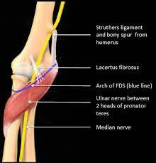
Fig 1
Etiology
Quick and repetitive grasping or pronation movements can cause pronator teres muscle hypertrophy and entrapment of the median nerve, especially in those individuals who have additional fibrous brands [6]. Pronator teres syndrome can develop after local trauma, compression by a schwannoma, and in patients undergoing anticoagulation therapy and renal dialysis [6-8]. Tight lacertus fibrosis also known as the bicipital aponeurosis can exacerbate the symptoms of pronator teres syndrome.
Epidemiology
Pronator teres syndrome is a rare condition with less than 1 per 100,000 cases annually. It can easily be overlooked and mistaken for the more prevalent carpal tunnel syndrome (CTS) [9,10]. It is common in the 5th decade. One study reported a higher prevalence of pronator teres syndrome in men [9].
Clinical Presentation
Pronator teres syndrome can manifest with pain in the volar aspect of the forearm. The pain is aggravated by resisted pronation of the forearm and flexion of the elbow. The tinel sign can be positive over the proximal edge of pronator teres [6]. The patient may complain of significant weakness. Wasting of the muscles innervated by the median nerve is rare in pronator teres syndrome. A mild weakness of flexor pollicis longus and abductor pollicis brevis is not uncommon, with some involvement of flexor digitorum profundus to the 2 and 3 digits and opponens pollicis. The pronator teres is usually spared because it receives innervation before the median nerve pierces it. Sensory loss is variable. Sensory loss involves the palm of the hand. It can mimic sensory loss associated with a carpal tunnel syndrome, including the thenar eminence, thumb, index, middle, and ring fingers. The phalen test is positive over the pronator teres muscle in 50% of cases [6].
Evaluation
Nerve conduction studies (NCS) should be done in pronator teres syndrome to rule out other neuropathies, but they seldom show any abnormalities [1]. In patients with severe and axonal symptoms, the sensory and motor amplitudes are reduced more than conduction velocities [6,9].
Electromyography (EMG) abnormalities occur in the flexor pollicis longus and flexor digitorum profundus to 2nd and 3rd digits, less often in the flexor digitorum superficialis and abductor pollicis brevis, and only rarely in the pronator teres. Abnormal electrodiagnostic findings are found in about 10% of the patients with pronator teres syndrome [7,11].
Differential Diagnosis
The differential diagnosis includes anterior interosseous nerve compressive neuropathy (Kiloh-Nevin's syndrome), carpal tunnel syndrome, and pronator teres strain.
Treatment
Conservative treatment includes rest, modification of activities that exacerbate the symptoms, splinting, nonsteroidal anti-inflammatory medications, and local injections of corticosteroids [2,12]. Conservative treatment should be conducted for at least 6 weeks. If 3 to 6 months of conservative treatment fails surgery is usually needed.
Surgical treatment is required when the patient is symptomatic, has one or more objective findings on physical examination, such as weakness or motor atrophy, and has abnormality on electrodiagnostic studies [12]. During surgery, exploration of the median nerve in the forearm is done with release of the pronator teres muscle as well as all other compressive structures such as the ligament of Struthers, lacertus fibrosis, and/or fascia of flexor digitorum superficialis [12].
Differential Diagnosis
Some authors combine pronator teres syndrome with the following proximal median nerve entrapment syndromes due to their similar clinical presentation [7,11]:
1. Ligament of Struthers entrapment - In this syndrome, the median nerve is compressed by a ligament running from the medial epicondyle to a bony spur on a distal medial humerus, which is present only in 1-2 % of the population. Supination of the forearm and extension of the elbow exacerbates pain in the forearm and paresthesias in the median-innervated digits. The radial pulse may also be diminished since the brachial artery runs next to the median nerve.
2. Hypertrophied lacertus fibrosis (bicipital aponeurosis) entrapment - This is the fascia that attaches the biceps to the ulna and it overlies the median nerve in the proximal forearm. The pain in the forearm and paresthesias in the median nerve innervated digits is exacerbated by resisted flexion of the elbow with the forearm in supination.
3. Sublimus bridge of the flexor digitorum superficialis muscle entrapment- The pain in the forearm and paresthesias in the median innervated digits is increased by resisted flexion of the proximal interphalangeal joint of the middle finger while other fingers are held in extension.
The most common median nerve entrapment syndrome is the carpal tunnel syndrome. Pronator teres syndrome should be distinguished from carpal tunnel syndrome as well as from anterior interosseous nerve syndrome, brachial plexus injury, and cervical radiculopathy.
The pronator teres syndrome has sensory loss in the entire median nerve distribution unlike the carpal tunnel syndrome, where sensation is spared over the thenar eminence because the palmar cutaneous branch leaves the median nerve proximal to the carpal tunnel but distal to the pronator teres muscle. Nocturnal paresthesia symptoms are usually absent in pronator teres syndrome [4]. The amplitude of the median nerve may decrease in the forearm, but the distal motor and sensory latencies are normal in pronator teres syndrome except when there is associated CTS [1,9,13,14].
Carpal tunnel syndrome is sometimes diagnosed, and the more proximal pronator teres syndrome is missed when both are present in the same limb [14]. Thus in patients with carpal tunnel syndrome, pronator teres syndrome should be ruled out if the patient is going for surgery [9,14].
In the anterior interosseous nerve syndrome, there is no sensory loss whereas in the pronator teres syndrome there is sensory loss and in some cases, pronator teres syndrome may have only mild paresthesias in the median nerve distribution. Clinically, in both the carpal tunnel syndrome and pronator teres syndrome, the patient presents with an inability to flex the distal phalanx of the thumb, index, and middle fingers and there is weakness of pronation.
In the case of cervical radiculopathy or brachial plexus injury, the examination will reveal a weakness in other muscles outside the median nerve territory. Neck pain radiating to the arm suggests the presence of cervical radiculopathy.
Prognosis
Good recovery occurs in patients who undergo pronator teres release for pronator teres syndrome. Patients return to light duty in about 3 weeks and regular duty in about 6 weeks [12]. Occupation therapy fastens recovery and is particularly useful in patients who have residual weakness.
Complications
Complications of surgical treatment are uncommon. In a study by Svernlövone et al [15] where 72 patients underwent surgery for proximal forearm nerve entrapment, no complications were recorded. The overall postoperative satisfaction rate was 59%. The possible complications from surgery include infection, seroma/hematoma formation, and nerve injury.
References
Lacey SH, Soldatis JJ. Bilateral pronator syndrome associated with anomalous heads of the pronator teres muscle: a case report. J Hand Surg Am. 1993 Mar;18(2):349-51.
Olewnik Ł, Podgórski M, Polguj M, Wysiadecki G, Topol M. Anatomical variations of the pronator teres muscle in a Central European population and its clinical significance. Anat Sci Int. 2018 Mar;93(2):299-306.
Vymazalová K, Vargová L, Joukal M. Variability of the pronator teres muscle and its clinical significance. Rom J Morphol Embryol. 2015;56(3):1127-35.
Nebot-Cegarra J, Perez-Berruezo J, Reina de la Torre F. Variations of the pronator teres muscle: predispositional role to median nerve entrapment. 1991-1992Arch Anat Histol Embryol. 74:35-45.
Hsiao CW, Shih JT, Hung ST. Concurrent carpal tunnel syndrome and pronator syndrome: A retrospective study of 21 cases. Orthop Traumatol Surg Res. 2017 Feb;103(1):101-103.
Hartz CR, Linscheid RL, Gramse RR, Daube JR. The pronator teres syndrome: compressive neuropathy of the median nerve. J Bone Joint Surg Am. 1981 Jul;63(6):885-90.
Afshar A. Pronator Syndrome Due to Schwannoma. J Hand Microsurg. 2015 Jun;7(1):119-22.
Farrell HF. Pain and the pronator teres syndrome. Bull Hosp Joint Dis. 1976 Apr;37(1):59-62.
Asheghan M, Hollisaz MT, Aghdam AS, Khatibiaghda A. The Prevalence of Pronator Teres among Patients with Carpal Tunnel Syndrome: Cross-sectional Study. Int J Biomed Sci. 2016 Sep;12(3):89-94.
Bair MR, Gross MT, Cooke JR, Hill CH. Differential Diagnosis and Intervention of Proximal Median Nerve Entrapment: A Resident's Case Problem. J Orthop Sports Phys Ther. 2016 Sep;46(9):800-8.
Olehnik WK, Manske PR, Szerzinski J. Median nerve compression in the proximal forearm. J Hand Surg Am. 1994 Jan;19(1):121-6.
Carter GT, Weiss MD. Diagnosis and Treatment of Work-Related Proximal Median and Radial Nerve Entrapment. Phys Med Rehabil Clin N Am. 2015 Aug;26(3):539-49.
Bridgeman C, Naidu S, Kothari MJ. Clinical and electrophysiological presentation of pronator syndrome. Electromyogr Clin Neurophysiol. 2007 Mar-Apr;47(2):89-92.
Bilecenoglu B, Uz A, Karalezli N. Possible anatomic structures causing entrapment neuropathies of the median nerve: an anatomic study. Acta Orthop Belg. 2005 Apr;71(2):169-76.
Svernlöv B, Nylander G, Adolfsson L. Patient-reported outcome of surgical treatment of nerve entrapments in the proximal forearm. Adv Orthop. 2011;2011:727689.
No comments:
Post a Comment