Developmental Dysplasia of the Hip (DDH)
Dr. KS Dhillon
Introduction
Abnormal hip development produces developmental dysplasia of the hip (DDH). It presents in infancy or early childhood with a spectrum ranging from dysplasia to dislocation of the hip joint.
It used to be referred to as congenital dislocation of the hip. Developmental dysplasia is more accurate as it explains the broad spectrum of hip joint abnormalities. The presentation can vary from minor hip instability to frank dislocation. The exact etiology is still not known. A combination of genetic, environmental, and mechanical factors play a role. In familial cases, many genetic loci have been identified. Early diagnosis and treatment is important to prevent residual DDH, which causes a limp, and or early osteoarthritis [1].
Etiology
The exact etiology of hip dysplasia is not known, but this condition does appear to be related to a number of different factors [2]. One such factor is racial background. In Native Americans and Laplanders, the prevalence of hip dysplasia is much higher (nearly 25-50 cases per 1000 persons) than in other races. The prevalence is very low in southern Chinese and black populations
[3-6].
An underlying genetic disposition appears to exist. The frequency of hip dysplasia is 10 times higher in children whose parents had DDH than in those whose parents did not [7].
Other factors related to DDH include intrauterine positioning and sex. Female sex, first-born child, and breech positioning are all associated with an increased prevalence of DDH. About 80% of individuals with DDH are female [8]. The rate of breech positioning in children with DDH is about 20% compared with 2-4% in the general population [9,10]. In some studies, the prevalence of DDH in females born in breech position has been estimated to be as high as 1 case in 15 persons [11].
Certain musculoskeletal disorders of intrauterine malpositioning or crowding, such as metatarsus adductus and torticollis, have been reported to be associated with DDH [12,13]. An increased prevalence of DDH is also reported to be associated with oligohydramnios [14]. The left hip is more commonly associated with DDH than the right hip. This is possible because of the common intrauterine position of the left hip against the mother's sacrum, which forces it into an adducted position [14]. Children in cultures where the mother swaddles the baby, forcing the infant's hips to be adducted, also have a higher rate of hip dysplasia [15].
Hip dysplasia can be associated with underlying neuromuscular disorders, such as myelomeningocele, arthrogryposis, cerebral palsy, and Larsen syndrome, though such cases are not usually considered DDH.
Epidemiology
The overall frequency of DDH is approximately 1 case per 1000 individuals. Barlow, however, believed that the incidence of hip instability during newborn examinations was as high as 1 case per 60 newborns [16]. According to a study by Barlow, more than 60% of newborns with hip instability became stable within the first week, and 88% became stable by age 2 months, leaving only 12% with residual hip instability.
The incidence of DDH varies from 0.06 in Africans to 76.1 per 1000 in Native Americans due to the combination of genetics and swaddling.
Unilateral involvement in 64% involves the left side due to in utero most frequent fetal positioning (left occipitoanterior). The left hip of the fetus in the uterus is adducted against the mother's lumbosacral spine [17,18].
Pathophysiology
The hip joint formation is highly dependent on the dynamic relationship between the femur and the acetabulum. Interference with proper contact between these two in utero or infancy can lead to DDH.
The lower limb buds develop around the 4th week. The chondroblasts aggregate to form the future bones of the hip joint. At six weeks the cartilage develops into femur diaphysis, precartilage into the future femoral head, which cannot be differentiated from the acetabulum. The blastemal cells form the trochanteric projection.
In the 7th week, the Interzone differentiates the sides of the hip joint. Proximally the acetabulum is formed as a shallow depression at 65 degrees. This later deepens to 180 degrees. The middle layer undergoes autolysis to form the joint space, synovial membrane, and ligamentum teres. By the 11th week, the hip joint is recognizable. In utero, the femoral head growth is faster than the acetabulum, which results in under-coverage of the femoral head. Hence any disturbance in the contact will lead to abnormal development.
Swaddling in an extreme position (immobile, extended, adducted hip) results in maligned contact between the acetabulum and femur and this prevents proper development of the hip. The acetabulum continues to grow up to age 5.
Maligned contact for a prolonged period leads to chronic changes such as hypertrophy of the capsule and ligament teres, and the formation of thickened acetabular edge, which further prevents the contact, and prevents the relocation of the femoral head [19].
History and Physical Examination
For mild hip instability, the clinical features vary. There is a limitation of abduction in the infant, asymmetric gait in the toddler, hip pain in adolescence, and osteoarthritis in the adult.
All newborns must be screened for DDH. Clinical examination is essential for the identification of DDH, especially for babies with risk factors. Hip instability/dislocation can be identified by Barlow and Ortolani maneuver.
To perform the Ortolani maneuver the infant is placed in the supine position, hip flexed to 90 degrees, and in neutral rotation. The clothes and diapers should be removed. This manoeuver will reduce the dislocated hip. The hip is held with the thumb on the inner aspect and the index and ring fingers on the greater trochanter. While applying anterior force on the greater trochanter, gently the hip is abducted. If the hip dislocates, a jerk or clunk would be felt. "Hip clicks" are not related to hip instability [20].
To perform the Barlow maneuver the child is placed in the same position as in the Ortlani maneuver. A posterior force is applied to the trochanter and the hip is adducted. A clunk or jerk will be felt if the hip can be dislocated. The maneuver is done gently as forceful adduction can cause instability [21]. The sensitivity of this maneuver when performed by experienced hands ranges from 87 to 97 percent and specificity varies from 98 to 99 % [22].
Asymmetry in the position and or the number of gluteal skin folds may be a clue for DDH. But asymmetry is normal in 27 % of infants.
Galeazzi (Allis) sign identifies real or apparent shortening of the femur, by comparing the knee height while the hip and knee are flexed and feet are flat on the table. The test will be positive when there is a dislocation of the hip.
Beyond the neonatal period, a limited range of hip movements could be a clue for the diagnosis of DDH. There is limited hip abduction of less than 75° or adduction of 30 degrees past the midline.
The Klisic test can be useful for the diagnosis of DDH. To perform the test the middle finger is placed over the greater trochanter and the index finger over the anterior superior iliac spine. When the hip is normal, the imaginary line between the two fingers points at or above the umbilicus. When the hip is dislocated, the trochanter is elevated, and the line projects inferior to the umbilicus.
Asymmetrical gait (Trendelenburg gait) due to abductor insufficiency, lumbar lordosis, toe walking, leg length discrepancies, and early hip osteoarthritis may indicate DDH.
Evaluation
There is a need for periodic surveillance for DDH throughout infancy. The goal is to prevent late presentation beyond six months.
The 2000 American Academy of Pediatrics clinical practice guideline recommends a hip ultrasound (US) at six weeks of age or an Xray of the hip at four months of age in girls with a positive family history of DDH or breech presentation in the third trimester. Universal screening is controversial. Most of the instability if found self-resolves [23].
In newborns when there are risk factors and the examination is normal an ultrasound is done at 6 weeks. This allows time for the resolution of physiologic immaturity and laxity.
When the examination is inconclusive or hip clicks are present a repeat examination is done in 2 to 4 weeks. If a positive Ortolani or Barlow test is present the newborn is referred to an orthopedic specialist with experience.
At 4 weeks to 4 months if the examination is inconclusive the child is referred to a specialist or a hip ultrasound is done at six weeks.
If the Barlow or Ortolani test is positive the child is referred to an orthopedic specialist with experience.
After 4 months clinical examination is limited as capsule laxity decreases. The Barlow and Ortolani maneuvers may not be positive. Limited hip abduction becomes the most useful screening method in older children.
Femoral head nuclei usually appear between 4 to 6 months so an X-ray is the preferred diagnostic tool. A normal pelvic radiograph at four months can reliably exclude DDH in children with risk factors.
The American Institute of Ultrasound in Medicine and the American college of radiology recommend an ultrasound of the hip [24]. The ultrasound is used to visualize acetabular dysplasia, hip dislocation, femoral head anatomy, ligament teres, and hip capsule. The femoral head coverage by acetabulum is determined. The minimum coverage should be at least 50% and the depth of the bony acetabulum is also determined. The Alpha angle of more than 60 degrees is considered normal.
The criteria for DDH has been established for static imaging by the Graf classification.
The Alpha angle is the angle between the bony acetabulum and ilium. The normal angle is more than 60 degrees. The Beta angle is the angle between the labrum and ileum. The normal angle is less than 55 degrees.
Based on these angles, DDH is classified into four types.
Graf Classification
Class Alpha angle Beta angle Description Treatment
I > 60° < 55° Normal None
II 43-60° 55-77° Delayed ossification Variable
III < 43° > 77° Subluxated Pavlik harness
IV Unmeasurable Unmeasurable Dislocated Pavlik Harness open and close reduction
AP X-ray of the pelvis is done and the following lines are measured (fig1).
Hilgenreiner's line: A horizontal line through the right and left triradiate cartilage. The head of the femur should be inferior to this line.
Perkin's line: Perpendicular line to Hilgenreiner's line through a focus on the lateral side of the acetabulum. The femoral head should be medial to this line.
Shenton's line: Smooth arc that connects the femoral neck to the superior margin of the obturator foramen. Any disruption indicates an abnormality.
Acetabular index: the intersection between Hilgenreiner's line and a line drawn tangential to the lateral ossific margin of the roof of the acetabulum. The normal index is less than 35 degrees at birth and less than 25 degrees at age one.
The center-edge angle of Wiberg: Formed by Perkin´s line and a line coming from the center of the femoral head to the lateral edge of the acetabulum. This measurement is reliable in patients older than 5 years old. Values should be more than 20 degrees.
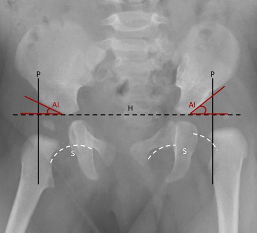
Fig 1.
Treatment
The aim of treatment is to provide an optimal environment for the growth of the femoral head and acetabulum. There is a need for a high index of suspicion and routine surveillance is needed to detect DDH and prevent complications.
There are various treatment modalities available to provide this optimal contact between the femoral head and the acetabulum. These include abduction splinting, closed reduction, and open reduction. The efficacy of
double diapering is close to zero.
At 0 to 4 weeks mild instability without a dislocatable hip can be kept under observation [25]. If the hip is dislocatable then an early referral to an orthopedic surgeon experienced in the treatment of DDH will be optimal. The application of Pavlik harness will be up to the orthopedic surgeon.
A study done by Larson JE et al concluded that waiting up to 30 days before initiation of treatment showed no significant difference in the outcome [26].
At 1 to 6 months abduction devices such as Pavlik harness, Von Rosen splint, Lausanne-developed abduction brace, Ilfeld orthosis, and Frejka pillow can be tried. The Pavlik harness is the widely used device for DDH. It consists of an anterior strap that flexes the hip at 90 degrees and prevents extension. The posterior strap prevents adduction. It is worn for about 23 hours per day for at least six weeks or until the hip is stable. An ultrasound of the hip is done every 3- 4 weeks to monitor the position of the femoral head. The success rate is about 90% for a Barlow's positive hip.
A high failure rate is seen with the following conditions [27]: Ortolani positive hip, initiation of treatment after seven weeks, multigravida, foot deformity, and male sex.
If the hips are not reduced by three weeks, semi-rigid, non-flexible abduction devices like Ilfed orthosis to keep the hip in an abducted position can be tried. In a study by Sankar et al. there was an 82% success rate with Ilfed orthosis after Pavlik harness failure [28].
For infants who are diagnosed with DDH at 6 to 18 months or patients who failed with abduction devices, closed reduction with a hip spica cast application is preferred. Under general anesthesia (GA), the hip is placed in 90-100 degrees flexion and 40-50 degrees abduction. The failure rate with close reduction is about 13.6 % [29].
The main complication of close reduction is avascular necrosis of the femoral head. A CT scan or MRI is needed to confirm the hip position. A study by Gornitzky AL et al [30] showed that an MRI with contrast can identify perfusion abnormalities following closed reduction, thus preventing AVN.
For children diagnosed with DDH after 18 months and in infants who failed closed reduction, open reduction is the preferred treatment. Open reduction can also correct anatomical abnormalities such as inverted labrum, neolimbus, pulvinar, and hypertrophied ligamentum trees. Capsulorrhaphy and release of tight iliopsoas tendon are also carried out. The two preferred approaches are the medial and anterior (Smith-Peterson) approaches. The medial is less invasive, but the anterior is classical and will help correct most of the anatomical abnormalities. A femoral shortening osteotomy can be done if needed. The main complication is AVN. After open reduction, a spica cast is applied, and reduction is confirmed with CT or MRI.
Children with shallow or vertical acetabulum can develop osteoarthritis due to edge loading. Children presenting with acetabular dysplasia up to age 5, without dislocation can be treated with part-time or full-time abduction orthosis.
After the age of 5 years, pelvic osteotomies (Salter, Pemberton, and Dega) can be done to increase anterior or anterolateral coverage. If the patient has an open triradiate cartilage center, a triple innominate osteotomy can be done.
Salvage pelvic osteotomies are indicated in patients more than 8 years old. A shelf procedure is indicated for patients more than 8 years old with a subluxed femoral head. An extra-articular buttress of bone is added to the lateral weight-bearing aspect of the acetabulum. A Chiari procedure is indicated for patients with inadequate femoral head coverage where a concentric reduction cannot be obtained. It medializes the acetabulum via iliac osteotomy.
Adolescent patients who present with hip pain and have a shallow acetabulum, the triradiate cartilage is close and there are no signs of hip degeneration, can be treated with a periacetabular osteotomy (PAO). Bernese PAO is a technique in which multiple cuts are made to modify and reorient acetabular cartilage while an intact posterior column is maintained [31].
Differential Diagnosis
The differential diagnosis includes the following:
A femoral neck fracture
Proximal femoral focal deficiency
Coxa vara
Residual effects of infective arthritis
Prognosis
The long-term outcome of treatment of DDH depends on the degree of acetabular dysplasia, the age when the diagnosis is made, the type of treatment given, and whether a concentric hip reduction was obtained.
Approximately 90 percent of neonatal hips with instability or mild dysplasia i.e Barlow-positive with an alpha angle of 50 to 60 and with 50% to 60 % of coverage, resolve spontaneously with normal functional and radiographic outcomes [32].
A 95% reduction is achieved with the Pavlik harness. Residual dysplasia occurs in about 20% of the children. If left untread over a prolonged period, there will be a gradual progression of functional disability and there can be accelerated osteoarthritis [16].
Complications
Failure to diagnose and treat DDH can lead to functional disability, hip pain, and early onset of osteoarthritis. The use of a pelvic harness can lead to several complications.
The most severe complication is Avascular necrosis of the femoral head. This complication can occur in upto 5% of the patients [33]. However, this complication can be prevented by proper fitting of the harness.
Femoral nerve palsy can be another complication. It should be suspected if the infant stops demonstrating spontaneous knee extension while in the Pavlik harness. The incidence is about 2.5 %. Most of them are associated with severe dysplasia or maintaining the hip flexion beyond 120 degrees. Femoral palsy usually resolves after the harness is removed.
Residual hip dysplasia is another complication. The child has to be followed up with x-rays bi-annually or annually till skeletal maturity. A normal X-ray at the age of two years of life indicates a good prognosis.
Other complications include skin irritation and knee subluxation. Complications following open or closed reduction include redislocation, osteonecrosis, infection, and stiffness.
Conclusion
Management of DDH requires early identification using appropriate technology such as ultrasound and x-rays and early referral to a specialist. There are clear guidelines for identifying and managing DDH.
The treatment depends on the age of the patient and the degree of dysplasia. Minor hip instability with Barlow test positive and Ortalani test negative recovers spontaneously in 90% of cases in the first two weeks of life. Continued surveillance by the primary care provider during infancy improves detection. Pavlik harness corrects 95% of DDH if applied before 6 months. Residual dysplasia can occur even after appropriate treatment, so annual follow-up is required until skeletal maturity.
Without early treatment, the child can develop a leg length discrepancy, limp, and limited hip abduction. DDH is the most common cause of early osteoarthritis in women under the age of 40 years.
Screening and identifying DDH before six months of age should be the goal to prevent long-term complications.
References
Kolb A, Chiari C, Schreiner M, Heisinger S, Willegger M, Rettl G, Windhager R. Development of an electronic navigation system for elimination of examiner-dependent factors in the ultrasound screening for developmental dysplasia of the hip in newborns. Sci Rep. 2020 Oct 02;10(1):16407.
Ziegler J, Thielemann F, Mayer-Athenstaedt C, Günther KP. [The natural history of developmental dysplasia of the hip. A meta-analysis of the published literature]. Orthopade. 2008 Jun. 37 (6):515-6, 518-24.
Skirving AP, Scadden WJ. The African neonatal hip and its immunity from congenital dislocation. J Bone Joint Surg Br. 1979 Aug. 61-B (3):339-41.
Getz B. The hip joint in Lapps and its bearing on the problem of congenital dislocation. Acta Orthop Scand Suppl. 1955. 18:1-81.
Hoaglund FT, Yau AC, Wong WL. Osteoarthritis of the hip and other joints in southern Chinese in Hong Kong. J Bone Joint Surg Am. 1973 Apr. 55 (3):545-57.
RABIN DL, BARNETT CR, ARNOLD WD, FREIBERGER RH, BROOKS G. UNTREATED CONGENITAL HIP DISEASE. A STUDY OF THE EPIDEMIOLOGY, NATURAL HISTORY, AND SOCIAL ASPECTS OF THE DISEASE IN A NAVAJO POPULATION. Am J Public Health Nations Health. 1965 Feb. 55:SUPPL:1-44.
Bjerkreim I, Arseth PH. Congenital dislocation of the hip in Norway. Late diagnosis CDH in the years 1970 to 1974. Acta Paediatr Scand. 1978 May. 67 (3):329-32.
Wilkinson JA. A post-natal survey for congenital displacement of the hip. J Bone Joint Surg Br. 1972 Feb. 54 (1):40-9.
Carter CO, Wilkinson JA. Genetic and environmental factors in the etiology of congenital dislocation of the hip. Clin Orthop Relat Res. 1964 Mar-Apr. 33:119-28.
Salter RB. Etiology, pathogenesis and possible prevention of congenital dislocation of the hip. Can Med Assoc J. 1968 May 18. 98 (20):933-45.
Ramsey PL, Lasser S, MacEwen GD. Congenital dislocation of the hip. Use of the Pavlik harness in the child during the first six months of life. J Bone Joint Surg Am. 1976 Oct. 58 (7):1000-4.
Kumar SJ, MacEwen GD. The incidence of hip dysplasia with metatarsus adductus. Clin Orthop Relat Res. 1982 Apr. 164:234-5.
Weiner DS. Congenital dislocation of the hip associated with congenital muscular torticollis. Clin Orthop Relat Res. 1976 Nov-Dec. 121:163-5.
Dunn PM. Perinatal observations on the etiology of congenital dislocation of the hip. Clin Orthop Relat Res. 1976 Sep. 119:11-22.
Kutlu A, Memik R, Mutlu M, Kutlu R, Arslan A. Congenital dislocation of the hip and its relation to swaddling used in Turkey. J Pediatr Orthop. 1992 Sep-Oct. 12 (5):598-602.
Barlow TG. Early diagnosis and treatment of congenital dislocation of the hip. J Bone Joint Surg Br. 1962. 44-B:292-301.
Loder RT, Skopelja EN. The epidemiology and demographics of hip dysplasia. ISRN Orthop. 2011;2011:238607.
Shaw BA, Segal LS., SECTION ON ORTHOPAEDICS. Evaluation and Referral for Developmental Dysplasia of the Hip in Infants. Pediatrics. 2016 Dec;138(6).
Litrenta J, Masrouha K, Wasterlain A, Castaneda P. Ultrasound Evaluation of Pediatric Orthopaedic Patients. J Am Acad Orthop Surg. 2020 Aug 15;28(16):e696-e705.
Bond CD, Hennrikus WL, DellaMaggiore ED. Prospective evaluation of newborn soft-tissue hip "clicks" with ultrasound. J Pediatr Orthop. 1997 Mar-Apr;17(2):199-201.
Jones DA. Neonatal hip stability and the Barlow test. A study in stillborn babies. J Bone Joint Surg Br. 1991 Mar;73(2):216-8.
Patel H., Canadian Task Force on Preventive Health Care. Preventive health care, 2001 update: screening and management of developmental dysplasia of the hip in newborns. CMAJ. 2001 Jun 12;164(12):1669-77.
Hauk L. Developmental Dysplasia of the Hip in Infants: A Clinical Report from the AAP on Evaluation and Referral. Am Fam Physician. 2017 Aug 01;96(3):196-197.
American Institute of Ultrasound in Medicine; American College of Radiology. AIUM practice guideline for the performance of an ultrasound examination for detection and assessment of developmental dysplasia of the hip. J Ultrasound Med. 2009 Jan;28(1):114-9.
Lorente Moltó FJ, Gregori AM, Casas LM, Perales VM. Three-year prospective study of developmental dysplasia of the hip at birth: should all dislocated or dislocatable hips be treated? J Pediatr Orthop. 2002 Sep-Oct;22(5):613-21.
Larson JE, Patel AR, Weatherford B, Janicki JA. Timing of Pavlik Harness Initiation: Can We Wait? J Pediatr Orthop. 2019 Aug;39(7):335-338.
Vadillo P, Encinas-Ullan CA, Moraleda L, Albiñana J. Results of the Pavlik harness when treating Ortolani-positive hips: predictors of failure and arthrographic findings. J Child Orthop. 2015 Aug;9(4): 249-53.
Sankar WN, Nduaguba A, Flynn JM. Ilfeld abduction orthosis is an effective second-line treatment after failure of Pavlik harness for infants with developmental dysplasia of the hip. J Bone Joint Surg Am. 2015 Feb 18;97(4):292-7.
Race C, Herring JA. Congenital dislocation of the hip: an evaluation of closed reduction. J Pediatr Orthop. 1983 May;3(2): 166-72.
Gornitzky AL, Georgiadis AG, Seeley MA, Horn BD, Sankar WN. Does Perfusion MRI After Closed Reduction of Developmental Dysplasia of the Hip Reduce the Incidence of Avascular Necrosis? Clin Orthop Relat Res. 2016 May;474(5):1153-65.
Kamath AF. Bernese periacetabular osteotomy for hip dysplasia: Surgical technique and indications. World J Orthop. 2016 May 18;7(5):280-6.
Stevenson DA, Mineau G, Kerber RA, Viskochil DH, Schaefer C, Roach JW. Familial predisposition to developmental dysplasia of the hip. J Pediatr Orthop. 2009 Jul-Aug;29(5):463-6.
Grill F, Bensahel H, Canadell J, Dungl P, Matasovic T, Vizkelety T. The Pavlik harness in the treatment of congenital dislocating hip: report on a multicenter study of the European Paediatric Orthopaedic Society. J Pediatr Orthop. 1988 Jan-Feb;8(1):1-8.
No comments:
Post a Comment