Spontaneous osteonecrosis of the Knee
Dr. KS Dhillon
Introduction
Spontaneous osteonecrosis of the knee (SONK) was first described in 1968. It is a distinct clinical entity that is a common cause of acute knee pain and swelling [1]. There is no consensus regarding the etiology of the condition [2]. In 94% of the cases, the disease is found in the medial femoral condyle. It can, however, also affect the lateral femoral condyle, the patella, and the proximal tibia [3]. The onset of the disease is nonspecific and insidious. Hence the diagnosis and treatment can be challenging. It can lead to subchondral collapse, secondary osteoarthritis, and the need for surgical treatment [4].
Etiology
The onset of spontaneous osteonecrosis of the knee is insidious and its cause is not identifiable. This makes it distinguishable from secondary osteonecrosis of the knee where the cause is known [5].
Some authors have proposed that the primary etiology of SONK is a subchondral insufficiency fracture that results in localized osteonecrosis [6]. There are other investigators who have corroborated these findings through small case series, adding evidence in favor of a traumatic etiology [7]. It is believed that these insufficiency fractures lead to the accumulation of fluid in the bone marrow, resulting in focal ischemia and bone necrosis [8]. Earlier theories suggested a vascular etiology as the cause of the condition [9]. The exact pathogenesis of the disease remains unknown.
Hussain et al [8] in 2019 published a systematic review that re-examined the proposed etiologies of spontaneous osteonecrosis of the knee. They noted a strong association between meniscal tears and SNOK. They found meniscal tears in 50 to 100% of patients with spontaneous osteonecrosis of the knee. The extent of medial meniscus extrusion correlated to the stage and volume of SONK lesions [10]. The authors believed that disruption of the posterior medial meniscus root increases tibiofemoral contact pressures which alters normal knee biomechanics, leading to subchondral insufficiency fractures seen in SONK [8].
Epidemiology
The most common form of knee osteonecrosis is spontaneous osteonecrosis of the knee [11]. It typically affects patients between the ages of 50 to 60 years and sometimes affects patients who are older than that [3]. It commonly affects individuals who are active, exercise regularly and have a history of osteoporosis or osteopenia [3]. Usually, women are more often affected than men. There is a correlation between decreased bone mineral density and the incidence of SONK in women over the age of 60. There is also a correlation between posterior root tears of the medial meniscus and the incidence of SONK [8,12]. The true prevalence may be greater than currently reported since many patients who present with advanced osteoarthritis may have had SONK that went unrecognized [2].
History and Physical Examination
Patients with SONK present with acute onset, severe unilateral knee pain. It is usually localized to the medial side of the joint. History of trauma is absent. Patients usually can recall exactly when the symptoms started [7]. Pain is present at night and at rest. Pain is common with weight bearing and can be quite debilitating. There is localized tenderness on palpation over the area affected area. Patients will usually have mild synovitis. Examination will show a small effusion with limitation of movements of the knee [5].
Evaluation
After a comprehensive history has been taken and physical examination performed imaging is the next step. In the early course of the disease, the radiographs are normal. In the later stage of the disease x rays of the knee will show flattening of the involved condyles (fig 1).
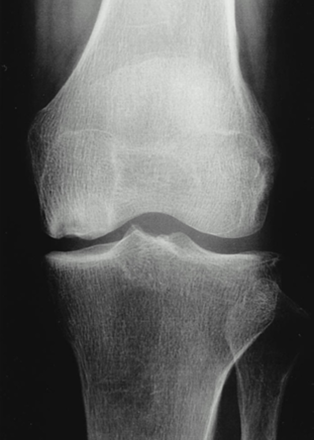
Fig 1.
In the early stage of the disease magnetic resonance imaging (MRI) is especially useful [11]. The MRI will show bone marrow edema localized to the medial femoral condyle, with extension into the inter-condylar notch. A subchondral crescent of a linear focus of low signal intensity can be seen (fig 2) [13]. The MRI can also show meniscal tears. Bone scintigraphy can show increased uptake in the area that is affected. However, the sensitivity and specificity of bone scintigraphy are lower than MRI [11].
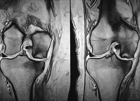
Fig 2.
Spontaneous osteonecrosis of the knee can be staged according to the Koshino classification that was described in 1979 [11]. It consists of four stages.
Stage I- A patient with knee symptoms but normal x-ray findings.
Stage II- A patients with flattening and subchondral radiolucencies without collapse.
Stage III- A patient with an extension of these radiolucencies with subchondral collapse.
Stage IV- A patient with further degenerative changes with osteosclerosis and osteophyte formation.
Treatment
Treatment of SONK depends on the extent and severity of the disease. In the initial stages of the disease, treatment is conservative, especially if the lesion is small [11]. Conservative treatment consists of lateral wedge insoles, multimodal analgesia, NSAIDs, protected weight-bearing, and bisphosphonates [13]. Yates et al. found complete resolution of symptoms in all 20 patients with stage I SONK treated conservatively [14].
Before subchondral collapse occurs, joint-preserving surgical procedures such as diagnostic and therapeutic arthroscopy, core decompression, and osteochondral autologous transplants can be carried out. Duany et al. reported an 87% success rate in preserving the knee joint using such techniques in patients with pre-collapse SONK at a mean follow-up of 40 months [15].
In patients with advanced disease, a high tibial osteotomy (HTO) can be carried out, especially in young patients. A unicompartmental knee replacement or a total knee replacement may be necessary when there is gross joint destruction. Both unicompartment arthroplasty and total knee replacement have shown favorable results as compared to those seen after total knee arthroplasty for osteoarthritis [16].
Differential Diagnosis
Since the onset of the disease is insidious and symptoms nonspecific it can be difficult to diagnose spontaneous osteonecrosis of the knee. Referred pain from concurrent hip disease as well as from intra-articular pathology has to be ruled out [17]. A good history and physical examination can help differentiate between SONK and other similar clinical entities. Differential diagnosis of SONK includes the following condition:
Osteochondritis dissecans
Transient osteoporosis
Shifting bone marrow edema
Secondary osteonecrosis
Bone contusion
Occult fractures
Prognosis
There is very little literature on the natural course and downstream consequences of SONK. The prognosis for the condition depends on the size of the lesion. Larger lesions increase the risk of osteoarthritis [18]. Jureus et al [4] reported on 40 patients with SONK with a mean follow-up of 15 years. They found that when 40% or more of the articular surface was involved OA was likely to develop. If the disease is detected in the early stages, non-operative treatment can be effective. If it is diagnosed late, patients are likely to develop end-stage osteoarthritis.
Complications
SONK is a progressive disease. The patient’s condition will continue to deteriorate if not diagnosed early and treated promptly. If it is diagnosed and treated early, patients can avoid risks and subsequent complications of surgery required for the treatment of advanced disease.
References
Ahlbäck S, Bauer GC, Bohne WH. Spontaneous osteonecrosis of the knee. Arthritis Rheum. 1968 Dec;11(6):705-33.
Mont MA, Marker DR, Zywiel MG, Carrino JA. Osteonecrosis of the knee and related conditions. J Am Acad Orthop Surg. 2011 Aug;19(8):482-94.
Zaremski JL, Vincent KR. Spontaneous Osteonecrosis of the Knee. Curr Sports Med Rep. 2016 Jul-Aug;15(4):228-9.
Juréus J, Lindstrand A, Geijer M, Robertsson O, Tägil M. The natural course of spontaneous osteonecrosis of the knee (SPONK): a 1- to 27-year follow-up of 40 patients. Acta Orthop. 2013 Aug;84(4):410-4.
Ecker ML, Lotke PA. Spontaneous Osteonecrosis of the Knee. J Am Acad Orthop Surg. 1994 May;2(3):173-178.
Yamamoto T, Bullough PG. Spontaneous osteonecrosis of the knee: the result of subchondral insufficiency fracture. J Bone Joint Surg Am. 2000 Jun;82(6):858-66.
Narváez JA, Narváez J, De Lama E, Sánchez A. Spontaneous osteonecrosis of the knee associated with tibial plateau and femoral condyle insufficiency stress fracture. Eur Radiol. 2003 Aug;13(8):1843-8.
Hussain ZB, Chahla J, Mandelbaum BR, Gomoll AH, LaPrade RF. The Role of Meniscal Tears in Spontaneous Osteonecrosis of the Knee: A Systematic Review of Suspected Etiology and a Call to Revisit Nomenclature. Am J Sports Med. 2019 Feb;47(2):501-507.
Jones JP. Alcoholism, hypercortisonism, fat embolism and osseous avascular necrosis. 1971. Clin Orthop Relat Res. 2001 Dec;(393):4-12.
Yasuda T, Ota S, Fujita S, Onishi E, Iwaki K, Yamamoto H. Association between medial meniscus extrusion and spontaneous osteonecrosis of the knee. Int J Rheum Dis. 2018 Dec;21(12):2104-2111.
Karim AR, Cherian JJ, Jauregui JJ, Pierce T, Mont MA. Osteonecrosis of the knee: review. Ann Transl Med. 2015 Jan;3(1):6.
Akamatsu Y, Mitsugi N, Hayashi T, Kobayashi H, Saito T. Low bone mineral density is associated with the onset of spontaneous osteonecrosis of the knee. Acta Orthop. 2012 Jun;83(3):249-55.
Yang WM, Zhao CQ, Lu ZY, Yang WY, Lin DK, Cao XW. Clinical Characteristics and Treatment of Spontaneous Osteonecrosis of Medial Tibial Plateau: A Retrospective Case Study. Chin Med J (Engl). 2018 Nov 05;131(21):2544-2550.
Yates PJ, Calder JD, Stranks GJ, Conn KS, Peppercorn D, Thomas NP. Early MRI diagnosis and non-surgical management of spontaneous osteonecrosis of the knee. Knee. 2007 Mar;14(2):112-6.
Duany NG, Zywiel MG, McGrath MS, Siddiqui JA, Jones LC, Bonutti PM, Mont MA. Joint-preserving surgical treatment of spontaneous osteonecrosis of the knee. Arch Orthop Trauma Surg. 2010 Jan;130(1):11-6.
Myers TG, Cui Q, Kuskowski M, Mihalko WM, Saleh KJ. Outcomes of total and unicompartmental knee arthroplasty for secondary and spontaneous osteonecrosis of the knee. J Bone Joint Surg Am. 2006 Nov;88 Suppl 3:76-82.
Mont MA, Baumgarten KM, Rifai A, Bluemke DA, Jones LC, Hungerford DS. Atraumatic osteonecrosis of the knee. J Bone Joint Surg Am. 2000 Sep;82(9):1279-90.
Lotke PA, Abend JA, Ecker ML. The treatment of osteonecrosis of the medial femoral condyle. Clin Orthop Relat Res. 1982 Nov-Dec;(171):109-16.
No comments:
Post a Comment